


Artificial intelligence in interventional cardiology: from procedural planning to intelligent cath lab ecosystems
 0
0 Abstract
Artificial intelligence (AI) is increasingly being evaluated across multiple stages of interventional cardiology, including pre-procedural planning, intraprocedural imaging and navigation, post-procedural monitoring, and decision-support systems. However, the maturity of evidence varies substantially across applications. computed tomography-derived and angiography-derived physiological assessment, selected automated image-analysis tools, and operator-controlled robotic assistance have accumulated prospective or real-world evidence in specific clinical settings, whereas digital twins, hybrid intravascular ultrasound-optical coherence tomography interpretation, generative simulations, autonomous robotic control, and large language model-based reasoning remain at different stages of validation. This review summarizes current AI applications across the interventional cardiovascular care pathway, critically appraises the strength of supporting evidence, and discusses translational barriers including dataset heterogeneity, workflow integration, cost-effectiveness, cybersecurity, accountability, regulatory oversight, and clinician trust. Rather than treating AI as a uniform technology, we propose an evidence-calibrated framework that distinguishes clinically implemented tools from investigational and conceptual platforms. Future adoption should depend not on algorithmic novelty alone, but on prospective validation, transparent evidence grading, reproducibility, regulatory clarity, and demonstrable improvements in patient-centered outcomes.
Keywords
INTRODUCTION
Multidimensional complexity challenges and cognitive limitations facing precision intervention
Interventional cardiology has evolved from balloon angioplasty and bare-metal stents to contemporary drug-eluting platforms, physiologic lesion assessment, transcatheter structural interventions, and increasingly complex catheter-based therapies. In parallel, Artificial intelligence (AI) has been widely discussed in cardiovascular and precision medicine, with previous reviews emphasizing its potential for image interpretation, risk prediction, workflow support, and clinical decision support[1-4]. Nevertheless, the clinical value of AI depends on the specific task, data source, validation design, regulatory pathway, and ability to improve outcomes rather than on computational performance alone.
Modern catheterization laboratories generate high-dimensional data from coronary angiography, intravascular ultrasound (IVUS), optical coherence tomography (OCT), fractional flow reserve (FFR), echocardiography, computed tomography (CT), electrocardiography (ECG), hemodynamic monitoring, laboratory tests, medications, and longitudinal electronic health records (EHRs). Conventional operator judgment remains indispensable, but integrating heterogeneous data under procedural time constraints can be affected by inter-observer variability, platform differences, and incomplete information. AI may therefore assist selected tasks by improving measurement reproducibility, extracting latent image or signal features, and supporting risk stratification[2-6].
Intergenerational evolution and leap of AI core technology in the cardiovascular field
Against the backdrop of these systemic bottlenecks, AI has emerged as a potentially useful computational engine. AI in cardiovascular medicine has evolved from early machine learning models based on structured clinical data to deep learning approaches enabling automated image analysis and three-dimensional reconstruction, significantly improving the efficiency and reproducibility of cardiovascular imaging[2].
More recently, emerging frameworks such as federated learning and large language models (LLMs) have further expanded AI applications by facilitating multi-institutional data integration and supporting complex clinical reasoning from unstructured data[4,5]. By enabling multi-agent collaboration, these models may expand AI from image-assistance utilities toward broader decision-support functions, although their reliability, safety, and clinical utility require task-specific validation[2].
Intention and core argument of the review: constructing a “full-lifecycle” intelligent intervention ecology
The integration of AI within interventional cardiology currently occupies a pivotal juncture, transitioning from technical proofs-of-concept to broad, real-world clinical implementation[2]. To comprehensively map this interdisciplinary evolution, this review synthesizes data from authoritative registries and contemporary clinical trials to conceptualize a full-lifecycle intelligent interventional ecosystem.
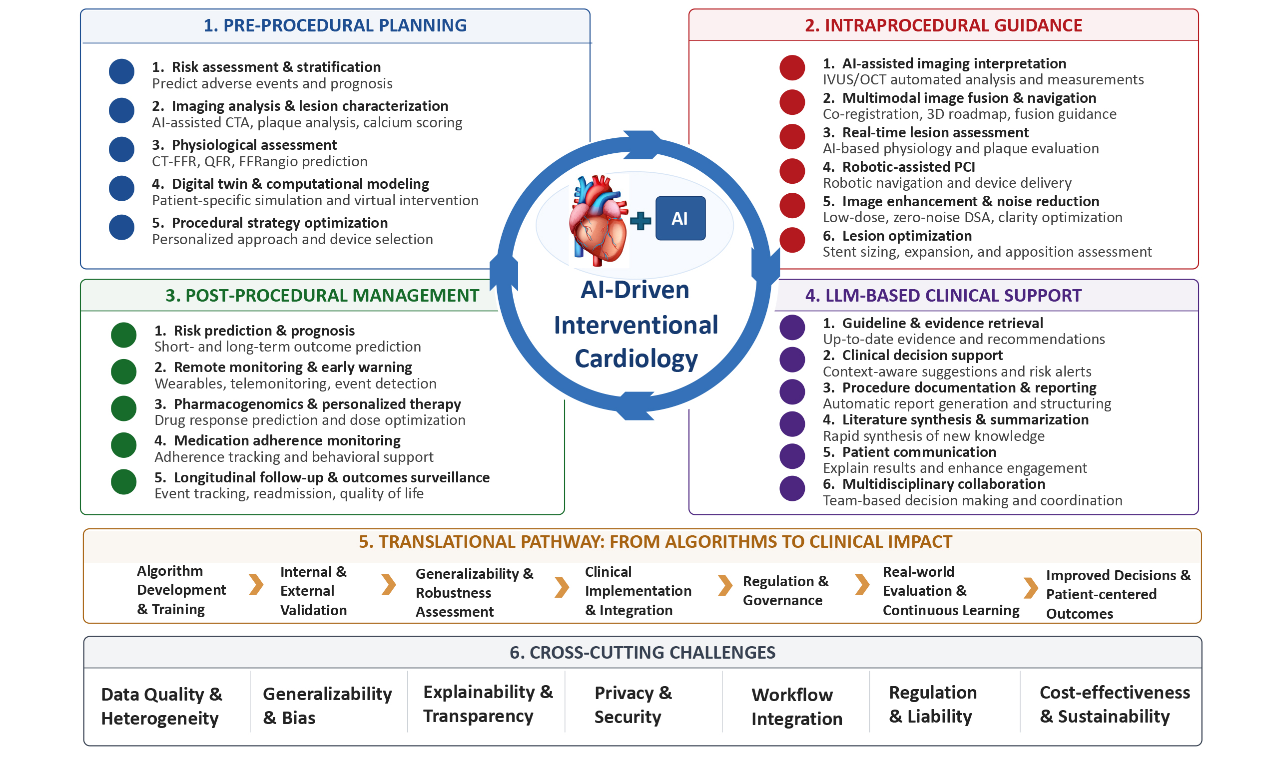
The scope of this review is intentionally organized according to the clinical pathway of interventional cardiovascular care rather than by algorithmic architecture. Coronary intervention, structural heart disease intervention, electrophysiology, robotics, intraprocedural imaging, wearable monitoring, pharmacogenomics, and LLM-based support are included because they represent distinct points at which AI may influence interventional decision-making: pre-procedural planning, intraprocedural guidance, post-procedural surveillance, and cognitive support. Importantly, these domains differ substantially in evidence maturity, regulatory pathways, workflow requirements, and clinical risk. Therefore, this review does not treat them as equivalent technologies, but evaluates them within an evidence hierarchy and translational framework.
PRE-PROCEDURAL PLANNING: FROM ANATOMICAL RECOGNITION TO FUNCTIONAL AND BIOMECHANICAL PREDICTION
Detailed, non-invasive preoperative planning is paramount for mitigating perioperative complications and optimizing long-term target vessel patency[6]. Digital twin technology facilitates the generation of patient-specific, virtual three-dimensional cardiac and hemodynamic models, enabling comprehensive strategy simulation and device matching prior to the physical procedure[6]. The foundation of this technology lies in transmuting imaging data into computable finite element models that synthesize tissue mechanical properties with device material characteristics, thereby facilitating the precise prediction of device-host interactions[7].
AI non-invasive image analysis and hemodynamic reconstruction for coronary intervention
Coronary CT angiography (CCTA) is an established non-invasive modality for evaluating coronary anatomy and plaque characteristics before invasive coronary procedures. Conventional CCTA interpretation, however, remains time-consuming and may be affected by inter-observer variability, particularly in the assessment of lesion severity, plaque composition, and high-risk plaque features[8]. AI-assisted segmentation and quantitative analysis may improve measurement reproducibility by automatically extracting the coronary tree, estimating lumen dimensions, and characterizing calcified, non-calcified, mixed, and low-attenuation plaque components[8,9]. These tools may also facilitate the identification of imaging features associated with higher-risk plaques, such as positive remodeling, low-attenuation plaque, spotty calcification, and the napkin-ring sign[8].
Beyond anatomical assessment, functional evaluation is essential for determining the clinical significance of intermediate coronary stenoses. CT-FFR and related computational approaches estimate lesion-specific ischemic potential from coronary anatomy and hemodynamic modeling[9]. Traditional computational fluid dynamics-based approaches may require complex segmentation and substantial computing resources, whereas machine learning-based approaches have been developed to accelerate pressure-gradient estimation by learning relationships between coronary geometry and invasive or simulated physiological measurements[9]. These methods may reduce calculation time and support non-invasive ischemia assessment, but their performance depends on image quality, model assumptions, acquisition protocols, and patient characteristics.
Clinical evidence supports the value of physiology-guided strategies in selected settings, although the cited technologies differ methodologically. The PLATFORM study reported favorable 1-year outcomes and lower healthcare costs with an fractional flow reserve derived from coronary computed tomography angiography (FFRCT)-guided diagnostic strategy in patients with suspected coronary disease[10]. The FAVOR III China (Functional Assessment by Various Flow Reconstructions III China) trial showed that angiography-derived quantitative flow ratio (QFR)-guided percutaneous coronary intervention (PCI) reduced 1-year major adverse cardiac events compared with angiography-guided PCI[11]. In patients with ST-segment elevation myocardial infarction and multivessel disease, the Compare-Acute trial demonstrated that invasive FFR-guided complete revascularization reduced the composite endpoint compared with culprit-lesion-only treatment[12]. Together, these studies support the broader principle that physiological assessment can refine revascularization decisions, but they should not be interpreted as evidence for a single AI-based non-invasive technology.
Several limitations should be emphasized. Diagnostic performance may vary across CT scanner generations, acquisition protocols, reconstruction algorithms, heart rate control, image noise, and motion artifacts. Severe coronary calcification remains a major challenge because blooming artifacts may impair lumen segmentation and pressure-gradient estimation[13]. In addition, computational fluid dynamics (CFD)-based FFRCT, machine learning-based CT-FFR, on-site algorithms, angiography-derived QFR, and invasive FFR-guided strategies are methodologically distinct and should be evaluated separately. Improved diagnostic classification or reduction in unnecessary angiography does not necessarily imply improvement in hard outcomes such as death or myocardial infarction. Finally, as shown by the ISCHEMIA trial, routine invasive treatment does not necessarily improve outcomes over optimal medical therapy in stable patients without appropriate clinical selection[13]. Therefore, real-world implementation of CT-derived and angiography-derived physiological tools requires careful attention to patient selection, reporting workflow, cost, interdepartmental collaboration, and prospective outcome validation.
3D modeling and finite element biomechanical simulation for structural heart disease intervention
Transcatheter interventions for structural heart disease require detailed pre-procedural assessment of complex three-dimensional anatomy, because device sizing, access planning, deployment depth, and interaction with calcified or deformable tissues may directly influence procedural safety and outcomes[6]. Patient-specific three-dimensional modeling and finite element simulation have therefore been explored as adjunctive tools for procedural planning. These approaches convert imaging data into computable anatomical and biomechanical models and may help estimate device-host interactions under different procedural scenarios[6,7].
In transcatheter aortic valve replacement (TAVR), CT-based three-dimensional assessment of the aortic root is central to valve sizing, annular evaluation, and prediction of procedure-related complications[14]. Finite element simulations may further support virtual valve deployment by estimating frame expansion, contact pressure, apposition to native calcification, and potential interaction with the conduction system[7,14]. In the PRECISE-TAVI study, CT-derived simulations showed potential value in predicting conduction disturbances requiring permanent pacemaker implantation and paravalvular leak in challenging anatomies[15]. These findings suggest that patient-specific simulation may provide additional information beyond anatomical measurements alone, particularly in anatomically complex cases.
Similar concepts have been applied to transcatheter mitral valve replacement (TMVR), where left ventricular outflow tract (LVOT) obstruction is a major procedural concern. CT-derived computer-aided simulations can estimate the predicted neo-LVOT area after device implantation. In a multicenter registry of 38 TMVR patients, predicted neo-LVOT area correlated with post-procedural CT measurements, and a predicted area ≤ 189.4 mm2 identified patients at high risk for LVOT obstruction with high sensitivity and specificity[16]. Such modeling may assist patient selection and planning of preventive strategies, including septal reduction therapy or anterior leaflet modification, when clinically appropriate.
For left atrial appendage occlusion, three-dimensional modeling may help characterize appendage morphology, assess landing-zone anatomy, and simulate device positioning[6]. In selected cases, virtual deployment of different occluder sizes or configurations may support procedural planning by estimating depth, coaxial alignment, residual leak risk, and device stability. However, these applications remain highly dependent on image quality, segmentation accuracy, modeling assumptions, and operator interpretation.
Overall, patient-specific biomechanical simulation remains an adjunctive planning approach rather than a validated determinant of treatment selection. Key limitations include assumptions regarding tissue material properties, boundary conditions, calcification stiffness, and device-tissue interaction. Many studies are based on small cohorts, retrospective validation, or simulation endpoints rather than prospective trials powered for clinical outcomes. Computational complexity, model standardization, vendor interoperability, and integration into time-sensitive procedural workflows also remain unresolved. Therefore, broader adoption of digital twin and biomechanical simulation technologies will require prospective validation, standardized modeling pipelines, and evidence that simulation-guided planning improves clinically meaningful outcomes.
Auxiliary mapping and substrate arrhythmogenic characteristic assessment for electrophysiological intervention
The persistent long-term recurrence rate following atrial fibrillation (AF) catheter ablation constitutes a paramount challenge within electrophysiology. By synthesizing time-frequency domain features extracted from surface ECGs with the three-dimensional spatial distribution of left atrial fibrosis derived from cardiac magnetic resonance imaging, multimodal machine learning frameworks can orchestrate patient-specific atrial arrhythmogenic substrate maps[17]. These deeply integrated models may support pre-procedural risk stratification for long-term recurrence, thereby directing operators on the clinical necessity of adjunctive ablation strategies—such as left atrial posterior wall linear ablation or complex fractionated atrial electrogram ablation—beyond conventional pulmonary vein isolation[17].
Furthermore, the primary prevention of sudden cardiac death (SCD) hinges critically on the preemptive identification of high-risk cohorts[2]. Traditional reliance on left ventricular ejection fraction (LVEF) thresholds is beset by significant diagnostic blind spots in both sensitivity and specificity, inadvertently excluding numerous high-risk patients from life-saving implantable cardioverter-defibrillator (ICD) therapy[2].
In the domain of myocardial strain analysis, AI has accomplished fully automated quantification. Regarding cardiomyopathy diagnostics, Goto et al.[18] deployed a multinational federated learning architecture that assimilated both ECGs and echocardiograms to screen for hypertrophic cardiomyopathy. This framework yielded cross-center C-statistics ranging from 0.90 to 0.96—vastly outperforming localized single-center models—thereby vividly illustrating the transformative potential of federated learning in the landscape of rare disease screening[18].
However, for electrophysiological applications, many AI models remain retrospective and endpoint definitions vary across studies. Prediction of AF recurrence or SCD risk should not be interpreted as evidence that AI-guided ablation or device implantation improves outcomes. Prospective trials are needed to determine whether model-guided substrate modification, ICD selection, or follow-up intensity changes clinical endpoints.
INTRAPROCEDURAL GUIDANCE: AI-ASSISTED IMAGING, NAVIGATION, AND ROBOTIC SUPPORT
For decades, the core of interventional cardiology has relied on two-dimensional X-ray fluoroscopic "silhouettes", which possess inherent limitations: tissue overlap, geometric distortion, and a fundamental inability to visualize soft tissue pathology within the vessel wall. As high-performance computing migrates to the surgical edge, AI applications within the catheterization laboratory are elevating procedural navigation to real-time, three-dimensional microscopic vision integrated with multimodal perception fusion[1].
AI real-time interpretation of intracavitary imaging and physical limit breakthrough of hybrid IVUS-OCT
In contemporary catheterization laboratories, IVUS and OCT are important adjuncts for guiding complex PCI[19]. However, their high frame rates and the need for manual measurement may limit rapid interpretation in urgent or complex procedures. AI-based algorithms can assist with automated plaque characterization, lumen and stent assessment, and detection of stent malapposition or edge dissection during image pullback[19].
Hybrid IVUS-OCT has been proposed to combine the complementary strengths of both modalities: the high axial resolution of OCT and the deeper tissue penetration of IVUS[1]. After the first-in-human report of a hybrid IVUS-OCT catheter in 2018[20], Bajaj et al. evaluated a deep learning classifier using hybrid IVUS-OCT pullbacks from 10 human cadaveric hearts with co-registered histology as the reference standard[21]. The hybrid model achieved an overall tissue classification accuracy of 86.7%, compared with 73.2% for IVUS, 66.6% for OCT, and 70.6% for blinded human experts[21]. For plaque phenotype detection, the hybrid model achieved a Cohen’s kappa of 0.60 and identified 68% of histologically confirmed thin-cap fibroatheromas[21].
Despite these encouraging findings, hybrid IVUS-OCT is not yet a routine clinical technology. Current evidence remains limited by small datasets, cadaveric or ex vivo validation, complex histological co-registration, and the absence of prospective outcome trials. Real-world adoption will depend on catheter cost, availability, compatibility with existing consoles, pullback workflow, contrast requirement, operator training, reimbursement, and whether additional tissue characterization improves PCI strategy or clinical outcomes beyond contemporary IVUS- or OCT-guided PCI.
Dual leap of multimodal real-time fusion navigation and zero-noise DSA technology
Algorithmic image enhancement is increasingly being explored to improve fluoroscopic and angiographic visualization in the catheterization laboratory, with AI-based image processing and multimodal visualization emerging as promising components of future image-guided interventions[4]. At Zhongshan Hospital, Fudan University, a zero-noise digital subtraction angiography (DSA) approach has been applied in complex coronary intervention, integrating radiographic hardware with software-based denoising and real-time image processing. This strategy aims to reduce image noise and improve vessel delineation, particularly in anatomically complex cases or procedures requiring careful contrast and radiation management[22]. However, its broader clinical value requires standardized validation of image quality, radiation dose, contrast use, procedural efficiency, and patient outcomes. Therefore, zero-noise DSA should currently be viewed as a workflow-supportive imaging technology rather than a validated replacement for established low-dose imaging protocols or operator judgment.
Sub-millimeter micro-distance operation of interventional surgical robots and mechanical circulatory monitoring
Interventional robotic systems have been introduced to improve procedural precision and reduce occupational radiation exposure and musculoskeletal strain for operators[4]. Current robotic PCI platforms should primarily be regarded as operator-controlled mechanical assistance systems rather than autonomous AI-guided intervention. By allowing the operator to manipulate guidewires, balloons, and stents from a shielded cockpit, these systems may improve ergonomic safety while maintaining physician control over procedural decision-making[4].
Clinical evidence for robotic PCI is emerging but remains mainly based on registries and observational comparisons. In an international multicenter real-world registry evaluating the second-generation CorPath GRX system, Mahmud et al. reported high technical and clinical success rates across several lesion subsets, including chronic total occlusions, bifurcations, calcified lesions, angulated lesions, and long lesions[23]. In a propensity score-matched analysis of 996 consecutive PCI patients, Patel et al. found that robotic PCI was associated with lower radiation exposure metrics, including air kerma and dose-area product, without significant increases in contrast use or fluoroscopy time, although procedural duration was modestly longer[24]. These findings suggest that robotic PCI may provide radiation-safety and ergonomic advantages in selected settings.
However, important implementation barriers remain. Robotic PCI requires dedicated equipment, disposable devices, staff training, compatibility with existing interventional tools, and protocols for rapid manual conversion during complications. Haptic feedback remains limited, and lesion selection, operator experience, and institutional workflow may influence outcomes. In addition, the current evidence base does not establish autonomous procedural decision-making or superiority in hard clinical endpoints. Future studies should clarify whether robotic assistance improves procedural safety, operator well-being, radiation exposure, workflow efficiency, and patient outcomes in broader real-world practice.
AI exclusive intervention strategy and device optimization for complex severe calcified lesions
Severe calcific lesions constitute a formidable barrier to optimal stent expansion and apposition during high-risk complex PCI[25]. Deep neural networks applied to multimodal intracavitary imaging have transitioned from rudimentary anatomical segmentation to the fully automated, three-dimensional topological mapping of plaque micro-phenotypes[25]. Contemporary data confirm that AI algorithms quantify calcific nodule volume, maximum thickness, longitudinal extent, and circumferential arc with pixel-level precision[25]. Leveraging these precise three-dimensional metrics, the system can prospectively recommend intravascular lithotripsy or rotational atherectomy over conventional cutting balloons upon detecting extremely severe calcification (defined as deep, > 0.5 mm thick, and encompassing > 270° of the circumference)[25]. This data-driven algorithmic triage substantially optimizes lesion preparation strategies.
Regarding CTOs, Rempakos et al.[3] engineered an XGBoost machine learning architecture to forecast guidewire crossing success, utilizing data from 12,136 cases within the PROGRESS CTO registry. The model achieved an area under the curve (AUC) of 0.78, significantly outperforming the traditional J-CTO score. Occlusion length, blunt stump morphology, and the presence of interventional collaterals emerged as the most heavily weighted predictors[3]. This predictive model has been translated into an online web application, delivering robust, real-time decision support for operators at the table. Prediction of guidewire crossing success should not be conflated with improved procedural success or safety unless tested in prospective implementation studies.
Workflow and evidence limitations
Despite these advances, AI-enabled intraprocedural guidance faces substantial workflow barriers. Hybrid intravascular imaging, robotic PCI, real-time image enhancement, and lesion-specific decision support require additional hardware, software integration, operator training, procedural standardization, and reimbursement pathways. Their use may prolong procedure preparation, increase equipment costs, and introduce new failure modes related to device compatibility, data latency, and system interoperability. Moreover, most evidence remains derived from observational cohorts, engineering validations, or selected expert centers. Therefore, successful cath lab implementation will require prospective workflow studies, usability testing, emergency fallback protocols, and demonstration of incremental clinical benefit beyond contemporary operator-guided practice.
POST-PROCEDURAL MANAGEMENT: DYNAMIC RISK PREDICTION, REMOTE MONITORING, AND IMPLEMENTATION CHALLENGES
Longitudinal data integration and dynamic risk prediction models
Discharge from the physical catheterization laboratory represents merely a single node within the continuous interventional care continuum. In an AI-driven smart healthcare ecosystem, advanced ensemble algorithms (e.g., CatBoost, random forests, or DeepSurv) continuously assimilate longitudinal EHRs, routine laboratory follow-ups, and outpatient pharmacy data via cloud infrastructure[1,2]. These ultra-high-dimensional prediction models non-linearly capture complex interactions among thousands of variables, dynamically forecasting post-PCI target lesion failure and bioprosthetic valve structural deterioration following TAVR[1,4]. Many post-PCI risk models have shown promising performance in retrospective studies, but their clinical utility remains to be established. Real-world implementation may be affected by changes in clinical practice and data quality, and prospective studies are needed to determine whether AI-assisted risk prediction improves patient outcomes.
Precision pharmacogenomics and wearable internet of things for remote monitoring
Contemporary AI pharmacology platforms synthesize individual pharmacogenomic sequencing data (e.g., CYP2C19 loss-of-function alleles) with expansive, real-world cardiovascular phenotype registries to construct personalized, machine learning-driven pharmacokinetic models. These algorithms can precisely titrate an individual’s micro-scale risks of heightened thrombogenicity against gastrointestinal bleeding, thereby tailoring the optimal potent P2Y12 inhibitor regimen and de-escalation strategy for each patient[1]. Concurrently, medical-grade wearables equipped with photoplethysmography generate massive streams of continuous physiological data[26]. Deep convolutional and recurrent neural networks demonstrate exceptional anti-interference feature extraction capabilities for non-stationary time-series data. Consequently, they detect not only occult AF but also sense signs of heart failure decompensation days in advance by tracking subtle autonomic nervous system fluctuations, thus facilitating the "subclinical-stage interruption" of disease progression[26]. Wearable-based detection of AF or heart failure decompensation may increase early diagnosis but can also generate false positives, patient anxiety, over-testing, and alert fatigue. Pharmacogenomic models require prospective evidence showing that algorithm-guided treatment selection improves ischemic and bleeding outcomes beyond guideline-based care.
FRONTIER BREAKTHROUGHS: THE MULTIDIMENSIONAL VALUE OF AI-ECG IN PREDICTING VALVULAR HEART DISEASE, AF, AND HEART FAILURE
Ultra-large-scale multimodal datasets and discrete-time survival model architectures
Historically, the conventional 12-lead ECG has been considered inherently limited in identifying valvular heart disease prior to the onset of overt acute electrical disturbances[27]. In late 2025, an international collaborative effort led by Liang Yixiu and Academician Ge Junbo published a landmark study in the European Heart Journal[27]. Utilizing an exceptionally large, real-world paired dataset from the Zhongshan Hospital database, the study also incorporated an independent external validation cohort of 34,214 patients from the Harvard-affiliated Beth Israel Deaconess Medical Center[27]. The algorithmic architecture innovatively embedded a discrete-time survival loss function into an advanced residual convolutional neural network, thereby capturing time-to-event dynamics to precisely characterize a patient’s disease progression probability over time[27].
Concurrently, AI-ECG has achieved an important development in AF screening. Attia et al. leveraged deep convolutional neural networks to identify patients with paroxysmal AF based solely on a standard 10-second 12-lead ECG recorded during normal sinus rhythm, achieving an AUC of 0.87[28]. This demonstrates that even in the absence of active AF episodes, the surface ECG harbors occult electrophysiological signatures decipherable by deep learning, yielding a highly scalable, non-invasive tool for population-level screening.
Predictive performance and additive clinical value
In forecasting future significant regurgitant valvular lesions, the internal test set yielded a C-index of 0.774 for future mitral regurgitation, 0.691 for aortic regurgitation, and 0.793 for tricuspid regurgitation[27]. Within Cox proportional hazards models adjusted for age, sex, and other baseline confounders, patients stratified into the AI-designated "highest risk quartile" exhibited a 7.6-fold higher risk of developing severe mitral regurgitation compared to the lowest quartile. The corresponding relative risks for significant aortic and tricuspid regurgitation were 3.8-fold and 9.9-fold, respectively[27]. Furthermore, additive value analyses revealed that integrating the AI-ECG system into a baseline predictive model (comprising solely demographic and echocardiographic variables) significantly augmented the net reclassification index for moderate-to-severe mitral regurgitation (0.353) and severe tricuspid regurgitation (0.630), suggesting incremental prognostic value uncaptured by conventional imaging paradigms[27].
Interpretability verification of biological mechanisms
To interrogate the algorithmic "black box", the research team deployed high-order interpretability techniques, such as variational autoencoders, confirming that AI-ECG predictions correlate robustly with subclinical cardiac chamber remodeling[27]. This mechanistic validation suggests that valvular degenerative pathologies are preceded by an extremely insidious mechanical expansion of the annular matrix. This microscopic remodeling projects subtle vector alterations onto the surface ECG, thereby substantially advancing the timeline for preventive clinical intervention[27].
Fairness considerations: performance disparities of AI-ECG across diverse populations
Kaur et al.[29] elucidated the presence of systematic biases within AI-ECG models for heart failure prediction, noting that algorithmic performance was significantly diminished in young Black women. However, by calibrating individualized probability thresholds based on race, age, and sex, the researchers achieved an 11-percentage-point improvement in the F1 score[29]. This finding underscores the importance of fairness evaluation before clinical deployment of AI models. However, incorporating demographic variables such as race and sex into predictive models also raises ethical considerations regarding fairness and equitable implementation in clinical practice.
AI-ECG models identify statistical patterns associated with future disease risk, but these associations should not be interpreted as causal mechanisms. Interpretability analyses may generate biologically plausible hypotheses, such as subclinical chamber remodeling, but they do not prove that the ECG features mediate disease development. Prospective studies are required to determine whether AI-ECG screening improves patient outcomes, avoids overdiagnosis, and remains calibrated across populations and ECG acquisition systems.
LARGE LANGUAGE MODELS AND COGNITIVE DECISION SUPPORT: EARLY PROMISE AND UNRESOLVED RISKS
LLM-based systems may support selected low-risk tasks in cardiovascular care, including clinical documentation, literature synthesis, guideline retrieval, patient education, and structured preparation of clinical summaries. In interventional cardiology, these tools may be particularly useful for organizing complex multimodal information before procedures, summarizing longitudinal records, and assisting communication between clinicians and patients. Evidence from patient-facing question-answering tasks suggests that LLM-generated responses can be rated favorably for quality and empathy in selected settings[30]. However, these findings should not be extrapolated to autonomous decision-making in high-stakes procedural care.
Institution-specific LLM-based clinical knowledge support systems may provide a platform for exploring domain-specific knowledge integration. Nevertheless, such systems should be considered investigational unless supported by independent benchmarking, external validation, hallucination testing, reproducibility assessment, version control, and peer-reviewed clinical evaluation. Current reviews emphasize that LLMs may be useful for documentation, education, and information retrieval, but their reliability as autonomous clinical agents remains limited by hallucination, temporal instability of training data, lack of transparent reasoning, and uncertain accountability[31].
In high-stakes interventional settings, LLM outputs should not be used as autonomous recommendations. Physician review, traceable source attribution, cybersecurity safeguards, privacy protection, and clear liability frameworks are essential prerequisites for deployment. Future studies should evaluate not only response accuracy, but also reproducibility across model versions, performance under adversarial or incomplete clinical inputs, integration into cath lab workflow, and the downstream effects of LLM-assisted decision support on patient safety, clinician workload, and medico-legal responsibility.
TRANSLATIONAL BARRIERS: EVIDENCE, GENERALIZABILITY, ETHICS, REGULATION, AND HEALTH ECONOMICS
Cracking data silos: the dual engine of federated learning and generative AI
Many commercialized AI models rely on centralized datasets, inadvertently engendering demographic homogenization and algorithmic bias[5]. Federated learning networks—exemplified by the PerFed-Cardio architecture—utilize a localized data retention paradigm (i.e., "data does not leave the domain"), uploading only de-identified, encrypted gradient information to a central server for aggregation. In small-sample, multicenter, and highly heterogeneous data environments, these networks achieve robust AUC scores comparable to traditional centralized learning while strictly adhering to rigorous medical privacy regulations[32]. Concurrently, generative AI, powered by conditional diffusion models and generative adversarial networks, can synthesize "high-fidelity digital twin images" encompassing rare pathological features. This fundamentally augments model robustness and generalizability for extreme or atypical clinical scenarios[33].
Federated learning mitigates but does not eliminate privacy and governance risks. Gradient leakage, site imbalance, inconsistent data quality, and unclear ownership of jointly trained models remain unresolved. In addition, cross-institutional AI development requires explicit agreements regarding data stewardship, intellectual property, audit rights, and responsibility for post-deployment monitoring.
Explainable AI and the cultivation of core clinical trust
Complex neural networks use high-dimensional, non-linear mappings and are often difficult to interpret directly[34]. Explainable AI (XAI) aims to improve transparency by identifying variables or image regions that contribute to model predictions[34]. For tabular data, SHapley Additive exPlanations (SHAP) and Local Interpretable Model-Agnostic Explanations (LIME) estimate feature contributions or local prediction patterns, whereas saliency maps and Gradient-weighted Class Activation Mapping (Grad-CAM) highlight influential regions on CCTA, OCT, or other images[34,35]. These tools may help clinicians assess whether model attention is anatomically plausible, but they do not provide definitive explanations of internal reasoning.
Explainability is also relevant to informed consent, device certification, clinical accountability, and liability assessment[36]. Patients should understand the extent of AI involvement, clinicians need interpretable outputs to judge recommendations, and regulators require traceable documentation[36]. Algorithmic fairness remains equally important. Obermeyer et al. showed that using healthcare cost as a proxy for health status led to systematic underestimation of Black patients’ needs, illustrating how biased labels can reproduce structural inequities[37].
However, XAI is not a complete solution. Interpretation methods may be unstable, misleading, or inconsistent across approaches[34]. Moreover, XAI should not be equated with causality: saliency maps, SHAP values, or LIME explanations may help inspect model behavior, but they do not prove that highlighted features are mechanistic drivers of disease, treatment response, or clinical benefit.
Dynamic regulatory frameworks for software as a medical device
International regulatory agencies are rapidly establishing dynamic, total-product-lifecycle oversight frameworks centered on the "software as a medical device" (SaMD) paradigm[38]. Future gold standards for regulatory approval will mandate not merely comprehensive audits of "good machine learning practices", but also the execution of large-scale, prospective, multicenter randomized controlled trials (RCTs). These trials must definitively confirm that AI-assisted modalities deliver tangible "hard endpoint" benefits, such as significant reductions in target lesion revascularization and long-term all-cause mortality[38]. Regulatory evaluation should include not only pre-market diagnostic performance but also post-market surveillance, drift monitoring, audit trails, human override mechanisms, cybersecurity testing, and predefined procedures for model updates. For semi-autonomous interventional systems, regulators will also need to define acceptable levels of automation, operator responsibility, and emergency fallback requirements.
Health economics and cost-effectiveness: assessing value in AI-enabled interventional care
To achieve widespread clinical implementation, disruptive medical AI technologies must withstand rigorous health economic evaluations[39]. Recent cost-effectiveness analyses demonstrate that employing a deep learning-based perivascular fat attenuation index (FAI) to quantify coronary inflammation, subsequently integrated with Markov modeling to project long-term outcomes, effectively guides intensified lipid-lowering therapies and substantially curtails unplanned readmissions[40]. Yielding an incremental cost-effectiveness ratio operating well below accepted clinical thresholds, such systems are not only economically viable but proactively conserve critical healthcare resources[39], accelerating the paradigm shift from "empirical medicine" to "high-value-driven medicine"[40].
The FAVOR III China trial provided direct empirical evidence of these health economic advantages, demonstrating that the QFR-guided cohort achieved significant event reduction while utilizing less contrast media and requiring shorter procedural durations[11]. Parallel evidence is found in classical analyses of coronary artery calcium (CAC) score-guided statin therapy. Pletcher et al. determined that while blanket treatment of intermediate-risk patients is superior when statins are inexpensive and have negligible quality-of-life impacts, CAC-guided therapy becomes highly cost-effective when pharmaceutical costs rise or quality of life is impacted[41]. The ORFAN study provided extensive clinical validation of the AI-Risk algorithm in patients lacking obstructive coronary artery disease (CAD)[42]. Among 40,091 consecutive CCTA patients monitored for a median of 2.7 years, individuals without obstructive CAD comprised 81.1% of the cohort, yet contributed 66.3% of major adverse cardiac events and 63.7% of cardiac deaths. The FAI score across any coronary artery independently predicted these events. The AI-Risk classification effectively stratified patients into low/medium-risk, high-risk, and very-high-risk cohorts, revealing an 8-year cardiac death hazard ratio (HR) of 6.75 for the very-high-risk vs. low/medium-risk groups. This robust dataset establishes a formidable clinical foundation for the utility of AI-Risk and underpins the aforementioned cost-effectiveness projections[40,42].
Health economic evaluation should account for capital expenditure, software licensing, cloud infrastructure, cybersecurity, staff training, maintenance, workflow delay, downstream testing after false-positive alerts, and opportunity costs. A tool that improves diagnostic efficiency may still be economically unattractive if it increases procedure time, requires additional staff, or fails to improve hard clinical outcomes. Cost-effectiveness should therefore be evaluated from both hospital and payer perspectives, ideally alongside prospective implementation trials.
From evidence to practice: key nodes in the translational pathway
Because AI applications in interventional cardiology differ markedly in maturity, they should not be discussed as a uniform category. Technologies such as CT-derived physiology, angiography-derived QFR[10,11], selected AI-assisted intravascular image-analysis tools[21], robotic PCI[23-24], and AI-ECG screening have accumulated prospective, registry-based, or large-scale validation evidence in selected clinical contexts[28,43]. However, the strength of evidence varies across these applications, and improved diagnostic accuracy, procedural efficiency, or risk prediction should not be assumed to translate automatically into improved hard clinical endpoints.
Other technologies, including patient-specific digital twin simulations[7], hybrid IVUS-OCT interpretation[21], generative synthetic data, and LLM-assisted clinical reasoning[31], remain earlier in the translational pathway[33]. These approaches are promising but still require independent external validation, standardized data pipelines, workflow evaluation, regulatory assessment, and prospective studies demonstrating clinical utility. In particular, tools intended to influence procedural decisions should be evaluated not only for technical performance, but also for safety, reproducibility, operator interaction, failure modes, and downstream effects on patient outcomes.
Translation from research prototypes to routine clinical use requires several coordinated steps. These include regulatory review, post-deployment monitoring, sustainable reimbursement, integration with EHRs and cath lab systems, clinician training, patient acceptance, cybersecurity safeguards, and clear medico-legal accountability[44-46]. For continuously learning or semi-autonomous systems, additional requirements include audit trails, predefined update procedures, human override mechanisms, and emergency fallback protocols. Therefore, the future implementation of AI in interventional cardiology should be guided by evidence level, clinical risk, workflow compatibility, and demonstrable benefit rather than by algorithmic novelty alone. A summary of the evidence hierarchy and translational readiness of representative AI technologies is provided in Table 1.
Evidence hierarchy and translational readiness of AI technologies in interventional cardiology
| Domain | Example technologies | Current evidence level | Translational maturity | Key limitations |
| Coronary physiology[10,11] | CT-FFR, QFR | Prospective studies/RCT-level evidence for selected tools | Relatively mature | Scanner dependence, calcification, algorithm heterogeneity, patient selection |
| Intravascular imaging[19,21] | AI-OCT, AI-IVUS | Image-validation studies; limited prospective evidence | Emerging | Image endpoints predominate; limited outcome trials |
| Hybrid IVUS-OCT[20,21] | Hybrid catheter plus DL tissue classification | Histology-correlated validation; small datasets | Investigational | Cost, availability, workflow, training, lack of prospective PCI outcome data |
| Structural digital twin[6,7] | TAVR/TMVR simulation | Simulation studies and small validation cohorts | Emerging/investigational | Tissue-property assumptions, computation, vendor interoperability, no hard-endpoint RCTs |
| Robotic PCI[22,23] | Operator-controlled robotic systems | Registries and observational comparisons | Selectively implemented | Cost, learning curve, procedural duration, limited autonomy |
| AI-ECG[26,27] | AF, HF, valvular disease prediction | Large retrospective/prospective validation for selected tasks | Variable | Bias, false positives, overdiagnosis, uncertain outcome benefit |
| Wearables[25] | AF/HF monitoring | Consumer and clinical validation studies | Implemented but heterogeneous | Alert fatigue, false positives, workflow burden |
| Pharmacogenomics[1] | P2Y12 selection support | Genotype-guided evidence exists; AI-specific evidence limited | Emerging | Need prospective AI-guided outcome trials |
| LLM/CDSS[29,30] | Documentation, guideline retrieval, clinical reasoning support | Benchmarks and proof-of-concept studies | Early/investigational | Hallucination, reproducibility, liability, privacy |
| Generative simulation[4,32] | Synthetic imaging, digital cohorts | Engineering proof-of-concept | Conceptual/early | Validation, bias amplification, regulatory uncertainty |
CONCLUSION
AI is increasingly being evaluated across multiple stages of interventional cardiovascular care, but its clinical readiness varies substantially by application. Technologies such as CT-derived physiology, angiography-derived physiology, selected image-analysis tools, and robotic assistance have entered clinical workflows or accumulated prospective evidence in specific settings. By contrast, digital twins, hybrid IVUS-OCT interpretation, generative simulations, autonomous robotic control, and LLM-based reasoning remain largely investigational and require independent validation before routine clinical use.
Future progress should be measured not by algorithmic novelty alone, but by demonstrable improvements in patient-centered outcomes, procedural safety, workflow efficiency, equity, and cost-effectiveness. Prospective multicenter studies, external validation across heterogeneous imaging platforms and populations, transparent evidence grading, regulatory harmonization, cybersecurity safeguards, and clear medico-legal accountability will be essential. A clinically useful AI ecosystem in interventional cardiology should therefore be human-supervised, evidence-calibrated, workflow-compatible, and ethically governed.
DECLARATIONS
Authors' contributions
Conceptualization, literature search, drafting of the manuscript, and preparation of tables: Liang Y
Conceptualization, critical evidence appraisal, manuscript revision, response to reviewers, and language polishing: Li Z
Supervision, conceptual guidance, critical revision of the manuscript, and final approval of the submitted version: Ge J
All authors read and approved the final manuscript.
Availability of data and materials
Not applicable.
AI and AI-assisted tools statement
During the preparation of this manuscript, ChatGPT (OpenAI, GPT-5.5 Thinking model, accessed in July 2026) was used solely for language polishing and editorial refinement. The tool did not influence the study design, data collection, analysis, interpretation, or the scientific content of the work. All authors take full responsibility for the accuracy, integrity, and final content of the manuscript.
Financial support and sponsorship
None.
Conflicts of interest
Ge J is a Senior Advisory Editor of Vessel Plus. Ge J was not involved in any steps of editorial processing, notably including reviewers' selection, manuscript handling, or decision-making, while the other authors have declared that they have no conflicts of interest.
Ethical approval and consent to participate
Not applicable.
Consent for publication
Not applicable.
Copyright
© The Author(s) 2026.
REFERENCES
1. Nzeako TR, Elendu C, Echefu G, Olanisa O, Kiladejo A, Bob-Manuel ED. Artificial intelligence in interventional cardiology: a review of its role in diagnosis, decision-making, and procedural precision. Ann Med Surg. 2025;87:5720-34.
2. Sardar P, Abbott JD, Kundu A, Aronow HD, Granada JF, Giri J. Impact of artificial intelligence on interventional cardiology: from decision-making aid to advanced interventional procedure assistance. JACC Cardiovasc Interv. 2019;12:1293-303.
3. Rempakos A, Alexandrou M, Mutlu D, et al. Predicting successful chronic total occlusion crossing with primary antegrade wiring using machine learning. JACC Cardiovasc Interv. 2024;17:1707-16.
4. Samant S, Bakhos JJ, Wu W, et al. Artificial intelligence, computational simulations, and extended reality in cardiovascular interventions. JACC Cardiovasc Interv. 2023;16:2479-97.
5. Ryu H, Lee M, Kim S, Kim JH, Yang H. Federated learning for cardiovascular disease prediction: a comparative review of biosignal- and EHR-based approaches. Healthcare. 2025;13:2811.
6. Skalidis I, Stalikas N, Collet C, et al. Digital twins and simulations in transcatheter coronary and structural heart interventions. Eur Heart J Digit Health. 2026;7:ztaf129.
7. de Jaegere P, Roccatello G, Prendergast BD, de Backer O, Van Mieghem NM, Rajani R. Patient-specific computer simulation for transcatheter cardiac interventions: what a clinician needs to know. Heart. 2019;105:s21-7.
8. Pinna A, Boi A, Mannelli L, et al. Machine learning for coronary plaque characterization: a multimodal review of OCT, IVUS, and CCTA. Diagnostics. 2025;15:1822.
9. Mastrodicasa D, Van Assen M, Huisman M, et al. Use of AI in cardiac CT and MRI: a scientific statement from the ESCR, EuSoMII, NASCI, SCCT, SCMR, SIIM, and RSNA. Radiology. 2025;314:e240516.
10. Douglas PS, De Bruyne B, Pontone G, et al. 1-year outcomes of FFRCT-guided care in patients with suspected coronary disease: the PLATFORM study. J Am Coll Cardiol. 2016;68:435-45.
11. Xu B, Tu S, Song L, et al. Angiographic quantitative flow ratio-guided coronary intervention (FAVOR III China): a multicentre, randomised, sham-controlled trial. Lancet. 2021;398:2149-59.
12. Smits PC, Abdel-Wahab M, Neumann F, et al. Fractional flow reserve-guided multivessel angioplasty in myocardial infarction. N Engl J Med. 2017;376:1234-44.
13. Maron DJ, Hochman JS, Reynolds HR, et al. Initial invasive or conservative strategy for stable coronary disease. N Engl J Med. 2020;382:1395-407.
14. Agarwal A, Ranard L, Vahl T, Khalique O. Advanced computer simulation based on cardiac imaging in planning of structural heart disease interventions. J Clin Med. 2025;14:6885.
15. Hokken TW, Wienemann H, Dargan J, et al. Clinical value of CT‐derived simulations of transcatheter‐aortic‐valve‐implantation in challenging anatomies the PRECISE‐TAVI trial. Catheter Cardiovasc Interv. 2023;102:1140-8.
16. Wang DD, Eng MH, Greenbaum AB, et al. Validating a prediction modeling tool for left ventricular outflow tract (LVOT) obstruction after transcatheter mitral valve replacement (TMVR). Catheter Cardiovasc Interv. 2017;92:379-87.
17. Tang S, Razeghi O, Kapoor R, et al. Machine learning-enabled multimodal fusion of intra-atrial and body surface signals in prediction of atrial fibrillation ablation outcomes. Circ Arrhythmia Electrophysiol. 2022;15:e010850.
18. Goto S, Solanki D, John JE, et al. Multinational federated learning approach to train ECG and echocardiogram models for hypertrophic cardiomyopathy detection. Circulation. 2022;146:755-69.
19. Chu M, Jia H, Gutiérrez-Chico JL, et al. Artificial intelligence and optical coherence tomography for the automatic characterisation of human atherosclerotic plaques. EuroIntervention. 2021;17:41-50.
20. Sheth TN, Pinilla-Echeverri N, Mehta SR, Courtney BK. First-in-human images of coronary atherosclerosis and coronary stents using a novel hybrid intravascular ultrasound and optical coherence tomographic catheter. JACC Cardiovasc Interv. 2018;11:2427-30.
21. Bajaj R, Huang X, Alves-Kotzev N, et al. Deep learning-based plaque characterization in hybrid IVUS-OCT images is superior to single-modality deep learning analysis and human experts: head-to-head comparison against histology. Cardiovasc Res. 2026;122:245-57.
22. Zhou J, Zhang Q, Lu H, et al. Ultra-low-dose noise-free technology to reduce radiation exposure during coronary angiography. Circ: Cardiovasc Interv. 2026;19:e016542.
23. Mahmud E, Madder RD, Wohns DH, et al. Robotic-assisted percutaneous coronary intervention: final results of the PRECISION and PRECISION GRX studies. J Soc Cardiovasc Angiogr Interv. 2025;4:100456.
24. Patel TM, Shah SC, Soni YY, et al. Comparison of robotic percutaneous coronary intervention with traditional percutaneous coronary intervention: a propensity score-matched analysis of a large cohort. Circ Cardiovasc Interv. 2020;13:e008888.
25. Mccarthy KJ, Larnard EA, Anderson CK, et al. Automated artificial intelligence mapping of coronary plaque calcification: a comparison with manual intravascular image analysis. J Clin Med. 2025;14:8166.
26. Gaoudam N, Sakhamudi SK, Kamal B, et al. Wearable devices and AI-driven remote monitoring in cardiovascular medicine: a narrative review. Cureus. 2025;17:e90208.
27. Liang Y, Sau A, Zeidaabadi B, et al. Artificial intelligence-enhanced electrocardiography to predict regurgitant valvular heart diseases: an international study. Eur Heart J. 2025;46:4823-37.
28. Attia ZI, Noseworthy PA, Lopez-Jimenez F, et al. An artificial intelligence-enabled ECG algorithm for the identification of patients with atrial fibrillation during sinus rhythm: a retrospective analysis of outcome prediction. Lancet. 2019;394:861-7.
29. Kaur D, Hughes JW, Rogers AJ, et al. Race, sex, and age disparities in the performance of ECG deep learning models predicting heart failure. Circ Heart Fail. 2024;17:e010879.
30. Ayers JW, Poliak A, Dredze M, et al. Comparing physician and artificial intelligence chatbot responses to patient questions posted to a public social media forum. JAMA Intern Med. 2023;183:589.
31. Thirunavukarasu AJ, Ting DSJ, Elangovan K, Gutierrez L, Tan TF, Ting DSW. Large language models in medicine. Nat Med. 2023;29:1930-40.
32. Linardos A, Kushibar K, Walsh S, Gkontra P, Lekadir K. Federated learning for multi-center imaging diagnostics: a simulation study in cardiovascular disease. Sci Rep. 2022;12:3551.
33. Williams MC, Macarthur JAL, Forsyth R, Petersen SE. New ways to use imaging data in cardiovascular research: survey of opinions on federated learning and synthetic data. Eur Heart J Imaging Methods Pract. 2025;3:qyaf012.
34. Abbas Q, Jeong W, Lee SW. Explainable AI in clinical decision support systems: a meta-analysis of methods, applications, and usability challenges. Healthcare. 2025;13:2154.
35. Haupt M, Maurer MH, Thomas RP. Explainable artificial intelligence in radiological cardiovascular imaging—a systematic review. Diagnostics. 2025;15:1399.
36. Amann J, Blasimme A, Vayena E, Frey D, Madai VI. Explainability for artificial intelligence in healthcare: a multidisciplinary perspective. BMC Med Inform Decis Mak. 2020;20:310.
37. Obermeyer Z, Powers B, Vogeli C, Mullainathan S. Dissecting racial bias in an algorithm used to manage the health of populations. Science. 2019;366:447-53.
38. Armoundas AA, Singh JP. Total product lifecycle regulatory considerations and recommendations for generative AI-enabled medical devices. Eur Heart J Digit Health. 2026;7:ztag019.
39. El Arab RA, Al Moosa OA. Systematic review of cost effectiveness and budget impact of artificial intelligence in healthcare. NPJ Digit Med. 2025;8:548.
40. Tsiachristas A, Chan K, Wahome E, et al. Cost-effectiveness of a novel AI technology to quantify coronary inflammation and cardiovascular risk in patients undergoing routine coronary computed tomography angiography. Eur Heart J Qual Care Clin Outcomes. 2025;11:434-44.
41. Pletcher MJ, Pignone M, Earnshaw S, et al. Using the coronary artery calcium score to guide statin therapy: a cost-effectiveness analysis. Circ Cardiovasc Qual Outcomes. 2014;7:276-84.
42. Chan K, Wahome E, Tsiachristas A, et al. Inflammatory risk and cardiovascular events in patients without obstructive coronary artery disease: the ORFAN multicentre, longitudinal cohort study. Lancet. 2024;403:2606-18.
43. Armoundas AA. The electrocardiogram reimagined: AI-ECG as a prognostic beacon for valvular heart disease. Eur Heart J. 2025;46:4838-41.
44. Liu X, Li S, Zhu Q, Xu S, Jin Q. Interpretable semi-federated learning for multimodal cardiac imaging and risk stratification: a privacy-preserving framework. J Imaging Inform Med. 2025;38:2105-25.
45. Sengupta A, Alexis SL, Kovacic JC, Tang GHL. Current challenges in TAVI: neo-commissural alignment to mimic more physiologic valve implantation. Vessel Plus. 2020;4:40.
Cite This Article
 Open Access
How to Cite
Download Citation
Export Citation File:
Type of Import
Tips on Downloading Citation
Citation Manager File Format
Type of Import
Direct Import: When the Direct Import option is selected (the default state), a dialogue box will give you the option to Save or Open the downloaded citation data. Choosing Open will either launch your citation manager or give you a choice of applications with which to use the metadata. The Save option saves the file locally for later use.
Indirect Import: When the Indirect Import option is selected, the metadata is displayed and may be copied and pasted as needed.
About This Article
Special Topic
Copyright

Data & Comments
Data
0






Comments
Comments must be written in English. Spam, offensive content, impersonation, and private information will not be permitted. If any comment is reported and identified as inappropriate content by OAE staff, the comment will be removed without notice. If you have any queries or need any help, please contact us at support@oaepublish.com.