


Case Report: Navigating anticoagulation for atrial fibrillation in a patient with cancer
 0
0 Abstract
Atrial fibrillation (AF) and cancer frequently coexist. Cancer-associated hypercoagulability may exacerbate thrombogenesis in AF, while active malignancy confers elevated risks of bleeding and all-cause mortality. Consequently, the coexistence of hypercoagulability and bleeding risk in patients with AF and cancer poses a distinct clinical challenge for optimizing anticoagulation management. However, the optimal anticoagulation strategy for patients with concomitant AF and cancer remains insufficiently validated. Furthermore, tumor heterogeneity complicates the implementation of a uniform antithrombotic regimen. We report the case of a patient with AF and ulcerative-protruding, moderately differentiated rectal adenocarcinoma who underwent radical surgery (Dixon procedure). Gastrointestinal endoscopy was performed as part of postoperative oncological surveillance. Perioperative anticoagulation was managed by balancing bleeding and thrombotic risks in accordance with current guidelines and consensus statements. The postoperative antithrombotic therapy was individualized, taking into account the dynamic nature of cancer-associated thrombotic risk and the bleeding risks associated with invasive procedures during oncological follow-up. This case offers valuable insights into the management of patients with AF and cancer, highlighting the importance of an individualized treatment approach.
Keywords
INTRODUCTION
Atrial fibrillation (AF) is the most common sustained arrhythmia in the general population and is associated with a substantial burden of morbidity and mortality[1]. A population-based study demonstrated that cancer patients have a 1.6-fold increased risk of developing AF compared to age- and sex-matched controls without cancer, suggesting that cancer is an independent risk factor for AF. The reported prevalence of AF in cancer patients varies widely, from 3.1% to 44%, while the prevalence of cancer among patients with AF can be as high as 25%[2-4]. A Danish cohort study demonstrated a higher incidence of cancer after new-onset AF, with a particularly elevated risk of colorectal cancer within 90 days after AF diagnosis (hazard ratio [HR] 3.35 in men, 5.91 in women)[5]. This association was further highlighted in a Dutch bidirectional cohort study[6], where, after excluding individuals with prior cancer or AF, the one-year incidence of cancer in the AF cohort was 2.54% vs. 1.80% in matched controls. Similarly, the one-year incidence of AF in the cancer cohort was 2.84% vs. 1.19% in matched controls. A military cohort study suggested that AF is often diagnosed prior to colon cancer[7]. Although the value of new-onset AF as an indicator of occult malignancy remains debated, it is undeniable that AF patients requiring colonoscopy screening bear a higher burden of colorectal cancer[8].
AF and cancer share numerous common risk factors, including advanced age, obesity, smoking, altered autonomic tone, and endocrine and metabolic disorders. In addition, systemic inflammation is a shared pathophysiological mechanism linking both conditions. Consequently, their close association is not unexpected[9-11]. Conventional thromboembolic risk scores, such as CHA2DS2-VASc and CHA2DS2-VA, do not account for the cancer-related prothrombotic state and may therefore underestimate stroke risk in this population[12,13]. Similarly, the HAS-BLED score does not fully capture the heightened bleeding tendency conferred by cancer and its treatments. Notably, when cancer is included as a covariate, it emerges as the strongest predictor of bleeding in these models[12,14].
Management is further complicated by the frequent need for invasive diagnostic or therapeutic procedures. Periprocedural interruption of anticoagulation, such as for endoscopic examination, can paradoxically increase both thromboembolic and bleeding risks. Currently, evidence guiding optimal periprocedural and long-term anticoagulation strategy in patients with concomitant AF and cancer remains limited[15]. Consequently, dynamic adjustment of the anticoagulation regimen based on the specific clinical context is paramount in these patients.
CASE REPORT
A 66-year-old male patient was admitted to our hospital with a 9-year history of paroxysmal palpitations, which had worsened and were accompanied by shortness of breath over the past 20 days. Upon admission, the patient’s vital signs were: body temperature 36.6 °C, blood pressure 118/68 mmHg. An electrocardiogram (ECG) demonstrated AF with T-wave inversion. A summary of the patient’s medical history and anticoagulation regimen is presented in Table 1. The patient's past medical history included hypertension, diabetes, gastroesophageal reflux disease (GERD), and a history of ischemic stroke documented in prior neurology records. Medications included nifedipine, irbesartan, bisoprolol, atorvastatin, metformin, and ilaprazole. Symptomatic treatment for knee joint pain was provided in the form of a loxoprofen sodium gel patch. Coronary angiography performed in 2021 [Figure 1] demonstrated triple-vessel disease with an 85% localized, smooth-surfaced luminal stenosis in the second obtuse marginal branch. The plaque showed no signs of rupture, ulceration, or acute thrombus formation. Distal coronary blood flow was preserved (Thrombolysis in Myocardial Infarction flow grade 3, TIMI grade 3).
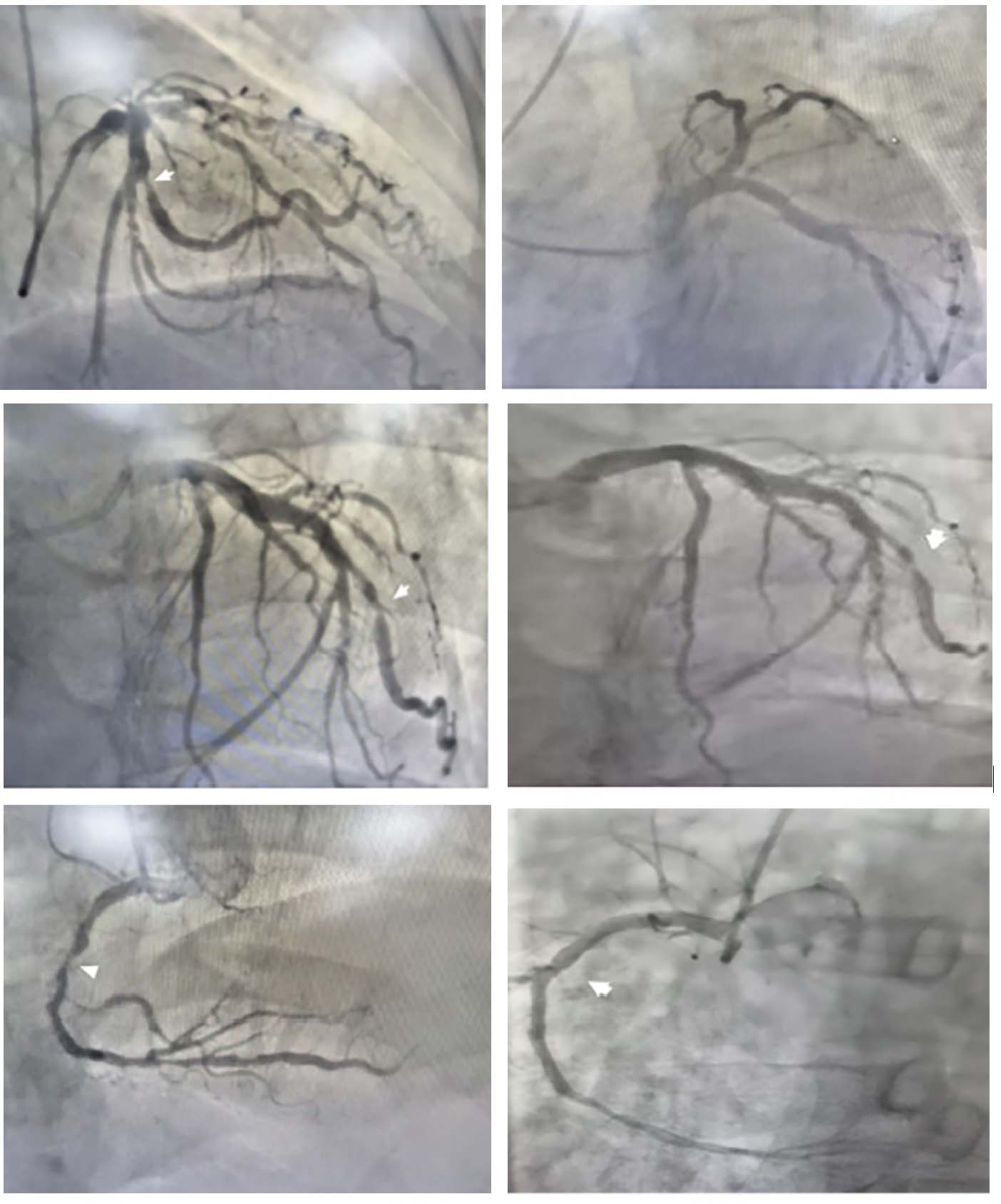
Figure 1. Coronary angiography. The site of coronary stenosis is indicated by the white arrow.
Medical history of the patient
| Date | Past medical history |
| 2016 | The patient presented with chest tightness and palpitations without an identifiable precipitating factor. Coronary computed tomography angiography (CTA) revealed triple-vessel disease, with the most severe luminal stenosis ranging from 55% to 60%. Accordingly, the patient was initiated on a medical regimen comprising aspirin, clopidogrel (for antiplatelet therapy), and isosorbide mononitrate sustained-release tablets |
| 2017 | Electrocardiogram (ECG) confirmed the presence of AF. Warfarin was subsequently initiated |
| 4 May 2018 | The anticoagulation therapy was switched to rivaroxaban due to warfarin intolerance |
| 9 Sep 2018 | The patient was scheduled for knee arthroplasty following an eight-year history of bilateral knee osteoarthritis that had worsened over the past two years. A preoperative stool occult blood test was positive. Subsequently, a colonoscopy revealed a moderately differentiated adenocarcinoma in the rectosigmoid colon. The patient underwent a 3D laparoscopic-assisted radical rectal resection (Dixon procedure) under general anesthesia. Postoperative pathological examination confirmed the diagnosis of an ulcerated, elevated-type, moderately differentiated adenocarcinoma of the rectum. Rivaroxaban was discontinued 48 h prior to surgery. Postoperative bridging therapy with enoxaparin sodium 0.6 mL (6000 anti-Xa IU) was initiated 24 h after surgery, following clinical assessment, and continued until discharge |
| 2 Nov 2018 | During a follow-up visit, rivaroxaban was replaced by dabigatran etexilate |
| 19 Oct 2021 | Coronary angiography revealed triple-vessel disease, including an 85% localized stenosis in the second obtuse marginal branch |
| 29 Oct 2025 | Positron emission tomography-computed tomography (PET-CT) revealed a focal hypermetabolic lesion in the stomach. Although the maximum standardized uptake value was below the threshold typically indicative of malignancy, the finding warranted clinical concern. Given the patient’s history of colorectal cancer, upper gastrointestinal endoscopy with biopsy was planned to rule out gastric metastasis or a second primary malignancy |
| 31 Oct 2025 | Prior to gastrointestinal endoscopy, the patient's anticoagulant was switched from oral dabigatran etexilate to subcutaneous enoxaparin sodium |
| 7 Nov 2025 | Following negative pathological findings (hyperplastic polyp, no malignant cells) from the gastrointestinal endoscopy, the bridging therapy with subcutaneous enoxaparin sodium was stopped. The oral anticoagulant regimen was subsequently transitioned to edoxaban |
Following admission, the patient underwent ambulatory blood pressure monitoring (ABPM), which revealed inadequate blood pressure control throughout the day. Blood pressure was intermittently above
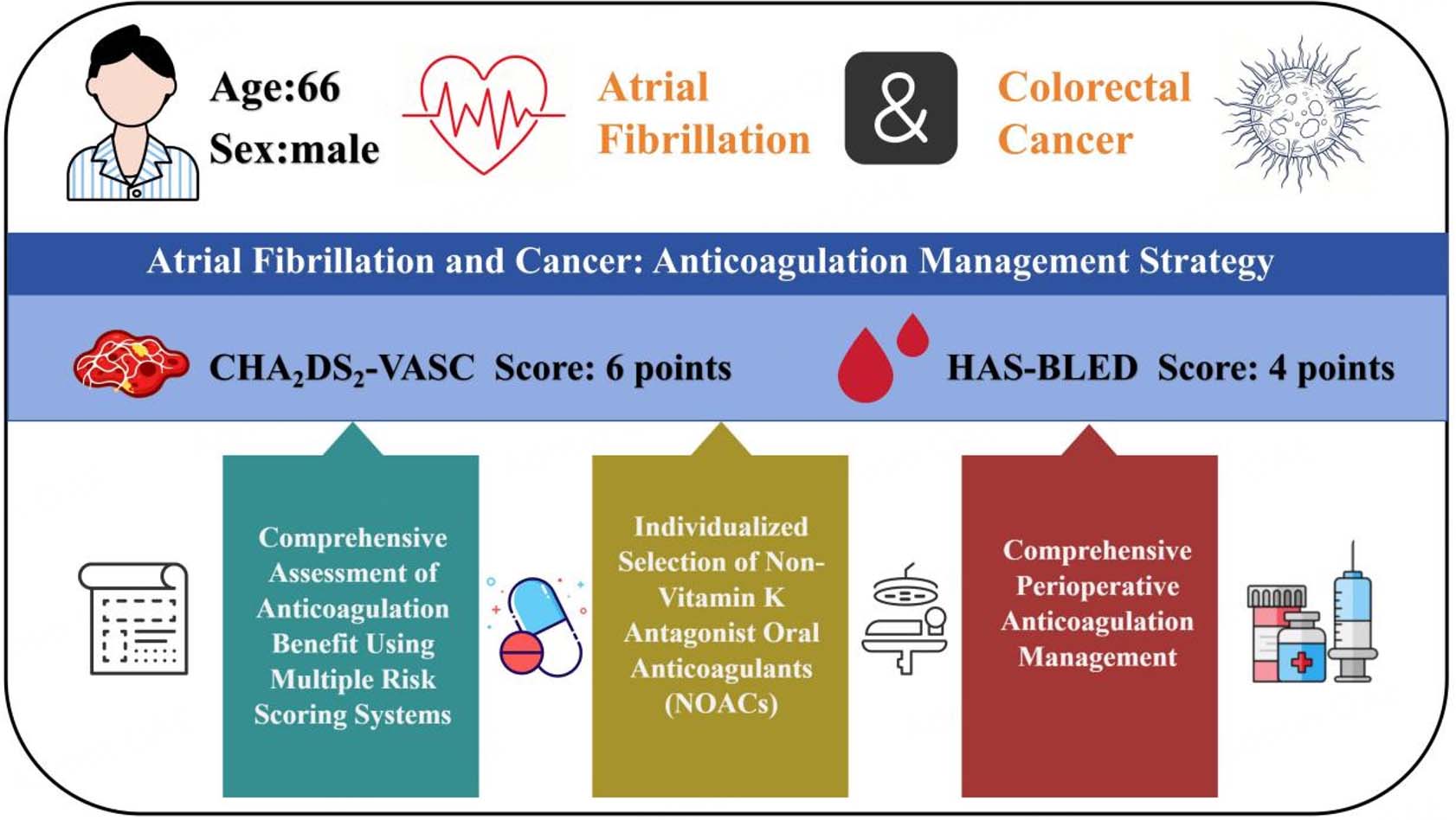
The patient's thrombotic and bleeding risks were assessed based on his clinical status. The patient's CHA2DS2-VASc score was 6, and his HAS-BLED score was 4 (Tables 2 and 3, respectively)[16]. The cancer-associated thrombosis risk was assessed [Table 4][17], and the patient's only factor meeting the score criteria of the Khorana risk score (KRS) and the Vienna Cancer and Thrombosis Study (CATS) score was a pre-chemotherapy leukocyte count of 12.9 × 10⁹/L. Figure 2 shows the adjustment process of antithrombotic therapy in this patient.
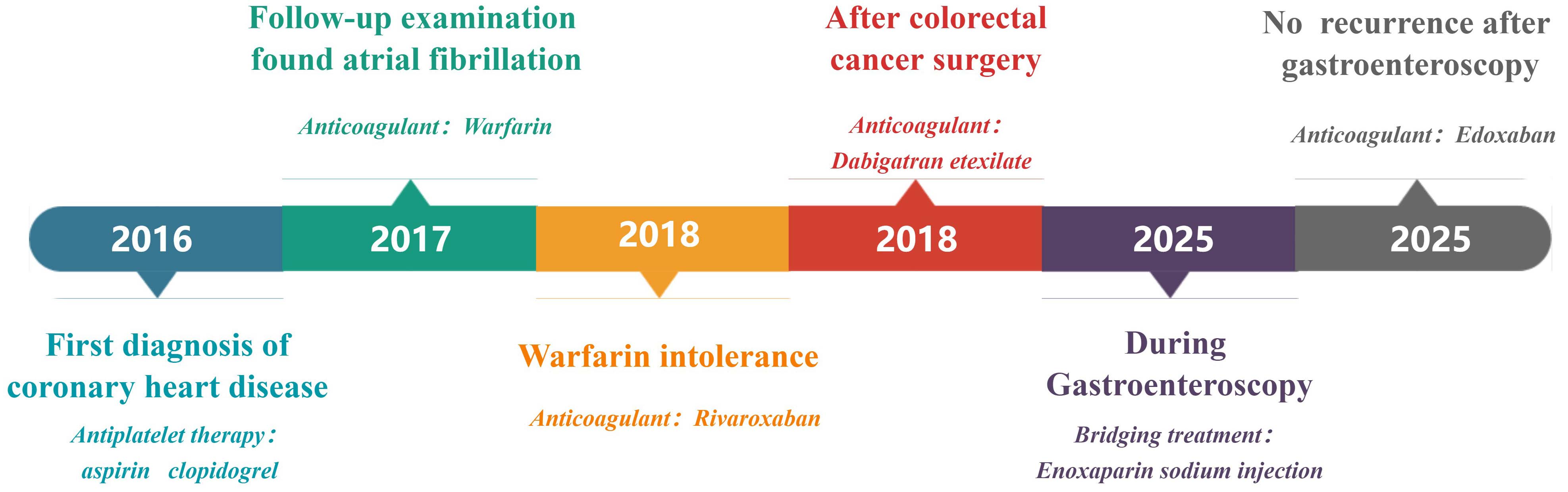
Figure 2. Schematic diagram of the patient's antithrombotic drug adjustment over time.
Scores for evaluation of thromboembolism risk. The numerical value indicates the points assigned to that specific risk factor. In the absence of parentheses, a factor is assigned a score of one point. Patient risk factors are highlighted in bold italicized text
| Score | Risk factors | Score points | Degree of stroke risk |
| CHA2DS2-VASc | Congestive heart failure (or left ventricular systolic dysfunction), Hypertension, Age ≥ 75 years (2), Diabetes mellitus, Prior stroke/TIA/Thromboembolism (2), Vascular disease, Age 65-74 years, Sex category (female) | 6 | High risk |
| CHADS2 | Congestive heart failure, Hypertension, Age ≥ 75 years, Diabetes mellitus, Stroke/TIA/Thromboembolism (2) | 4 | High risk |
| ATRIA | Age (prior stroke or not)*, Female, Diabetes mellitus, Congestive heart failure, Hypertension, Proteinuria, eGFR < 45 mL/min/1.73 m2 or ESRD | 9 | High risk |
Scores for evaluation of bleeding risk. The numerical value in parentheses indicates the points assigned to that specific risk factor. In the absence of parentheses, a factor is assigned a score of one point. Patient risk factors are highlighted in bold and italicized text
| Score | Risk factors | Score points | Degree of bleeding risk |
| HAS-BLED | Hypertension (uncontrolled), Abnormal liver and kidney function, Stroke, Bleeding, Labile INR, Age ≥ 65 years, Drugs/Alcohol, | 4 | High risk |
| ORBIT | Older age ≥ 75 years, Reduced hemoglobin/Anemia (2) Bleeding history (2) Insufficient kidney function (eGFR < 60 mL/min/1.73 m2), Treatment with Antiplatelet | 1 | Low risk |
| ATRIA | Anemia (3), Severe renal disease (eGFR < 30 mL/min/ 1.73 m2 or dialysis-dependent) (3), Age ≥ 75 years (2), Prior bleeding, Hypertension (diagnosed) | 1 | Low risk |
| HEMORR2HAGES | Hepatic or renal disease, Alcohol abuse, Malignancy, Age ≥ 75 years, Reduced platelet count or function, Rebleeding history (2), Hypertension (uncontrolled), Anemia, Genetic factors, Excessive fall risk, Stroke | 3 | Intermediate risk |
Cancer-associated thrombosis score at the time of initial rectal cancer diagnosis in 2018, prior to adjuvant chemotherapy. The numerical value in parentheses indicates the points assigned to that specific risk factor. In the absence of parentheses, a factor is assigned a score of one point. Patient risk factors are highlighted in bold and italicized text
| Score | Patient characteristics | Score points | Degree of thrombosis risk |
| Khorana risk score (KRS) | Pancreatic or gastric cancer (very high-risk tumors) (2), Lung, gynecological, lymphoma, bladder, or testicular (high-risk tumors), Prechemotherapy hemoglobin < 110 g/L or use of erythropoietin, Prechemotherapy leucocyte count > 11 × 109/L, Prechemotherapy platelet count > 350 × 10 9/L /, Body mass index ≥ 35 kg/m2 | 1 | Intermediate risk (High risk ≥3, Intermediate risk 1-2, Low risk 0) |
| Vienna-CATS score | Pancreatic or gastric cancer (very high-risk tumors) (2), Lung, gynecological, lymphoma, bladder, or testicular (high-risk tumors), Prechemotherapy hemoglobin < 110 g/L or use of erythropoietin, Prechemotherapy leucocyte count > 11 × 109/L, Prechemotherapy platelet count > 350 × 109/L, Body mass index ≥ 35 kg/m2, D-dimer > 1.44 μg/L, Soluble P-selectin* > 53.1 ng/L | 1 | Intermediate risk (High risk ≥ 3, Intermediate risk 1-2, Low risk 0) |
| COMPASS-CAT score | Prechemotherapy platelet count > 350 × 10 9/L (2), Previous venous thromboembolism, Metastatic disease/advanced (2), Time since cancer diagnosis < 6 months (4), Central venous catheter (3), Anti-hormonal therapy for women with hormone receptor-positive breast cancer or on anthracycline (6), Cardiovascular risk factors/comorbidities (≥ 2 of the following: personal history of peripheral arterial disease, ischemic stroke, coronary artery disease, hypertension, hyperlipidemia, diabetes mellitus, obesity) (5), Hospitalization (5) | 17 | High risk (Low/Intermediate risk ≤ 6, High risk > 7) |
Laboratory tests revealed the following: a B-type natriuretic peptide level of 657 pg/mL, a prolonged thrombin time of 18.2 s, a fibrinogen level of 1.78 g/L, and a positive initial fecal occult blood test. A complete blood count revealed a hemoglobin level of 138 g/L (reference range, 130-175 g/L) and a platelet count of 208 × 109/L (reference range, 125-350 × 109/L), both of which were within normal limits. Liver, renal, and thyroid function tests showed no significant abnormalities. Cardiopulmonary function, assessed via a six-minute walk test, was graded as Class II according to the American Thoracic Society guidelines. Transthoracic echocardiography demonstrated dilation of the aortic sinus and ascending aorta, biatrial enlargement, interventricular septal thickening, and mild mitral regurgitation [Figure 3]. Left ventricular wall motion and overall systolic function were preserved.
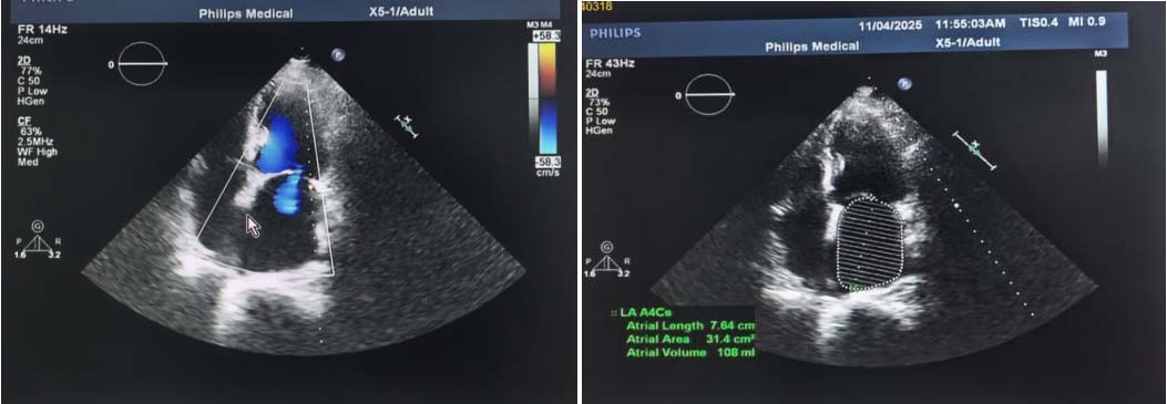
Figure 3. Transthoracic echocardiography performed at admission on 29 October 2025.
Following admission, the patient was prescribed a low-salt, low-fat diabetic diet. The pharmacological regimen was adjusted as follows: irbesartan 0.15 g once daily, benidipine 4 mg once daily, bisoprolol 2.5 mg once daily, ilaprazole 10 mg once daily, nicorandil 5 mg three times daily, sitagliptin phosphate 100 mg once daily, metformin 0.5 g three times daily, and atorvastatin 20 mg once daily. Notably, dabigatran etexilate
During hospitalization, the initial positive fecal occult blood test was attributed to a potential post-endoscopic procedure complication. A repeat fecal test two days later yielded a negative result. After three days of intermittent oxygen therapy and pharmacological management, the patient reported resolution of dyspnea and palpitations. Based on the negative gastric pathology findings confirming the absence of distal metastasis, the anticoagulant was transitioned to oral edoxaban 60 mg once daily. Following one week of observation without bleeding complications such as hematochezia, the patient was discharged.
After discharge, two telephone follow-ups were conducted with the patient: one on December 31, 2025, and another in mid-April 2026. During the first follow-up, the patient reported good medication adherence, having strictly followed the prescribed anticoagulation regimen without any missed or incorrect doses. No bleeding events (such as gingival bleeding, hematuria, or melena) or thromboembolic symptoms (such as chest pain, chest tightness, dyspnea, or lower extremity edema) were reported. The patient stated that he felt well overall and subjectively noted that the once-daily edoxaban regimen was more convenient compared to the previous twice-daily dabigatran etexilate regimen. During the second follow-up, the patient was residing in another city and was unable to return to Beijing; therefore the relevant original data (e.g., laboratory reports) could not be obtained. However, the patient had undergone basic laboratory testing at a local hospital and was informed by the attending physician that no abnormal findings of concern were identified. His condition remained consistent with that at the first follow-up: no bleeding or thromboembolic events had occurred, and his overall clinical status was stable.
DISCUSSION
Bleeding in patients with AF receiving anticoagulation therapy may be the initial manifestation of an occult malignancy. A cohort study of 119,480 participants identified a significant association between the site of malignancy and the origin of bleeding, with the strongest correlation observed for gastrointestinal cancers (HR = 15.4)[18]. Furthermore, in patients with AF diagnosed with cancer on direct oral anticoagulant (DOAC) therapy, a baseline bleeding risk score does not diminish the value of a cancer diagnosis as a strong predictor of major bleeding. The gastrointestinal tract is the most common bleeding site, with a reported incidence of 32.3%[19]. Therefore, clinicians should maintain a high index of suspicion for malignancy in all patients with AF receiving anticoagulation therapy, irrespective of their bleeding history. In the event of bleeding, a comprehensive evaluation based on the characteristics and site of hemorrhage is warranted to assess the possibility of a primary or metastatic malignancy.
Given the 4- to 7-fold increased risk of venous thromboembolism (VTE) and the 2-fold increased risk of bleeding during anticoagulation in patients with AF, developing individualized anticoagulation strategies that can be dynamically adapted to their evolving clinical status is particularly crucial[1]. In the present case, warfarin was initially prescribed by a general practitioner for a patient diagnosed with AF without known cancer. However, during follow-up at a tertiary hospital, the anticoagulant was switched to rivaroxaban by a cardiologist due to patient-reported intolerance to warfarin. This case highlights a cautious attitude toward DOACs among some Chinese physicians at that time, despite the 2014 American Heart Association, American College of Cardiology, and Heart Rhythm Society (AHA/ACC/HRS) guideline having already listed them as Class I recommendation[20]. Following a three-dimensional laparoscopic-assisted radical rectal resection (Dixon procedure) in 2018, the patient received dabigatran etexilate 110 mg twice daily for anticoagulation during chemoradiotherapy with oxaliplatin and capecitabine. Although observational and animal studies have suggested that vitamin K antagonists (VKAs) might inhibit AXL receptor signaling, enhance natural killer cell antitumor activity, and potentially reduce cancer risk, a large-scale study of 39,989 patients (31,200 on VKAs vs. 8,789 on DOACs) yielded the opposite conclusion. Compared to DOACs, VKAs demonstrated no superior cancer-preventive effect; instead, they were associated with a slightly increased overall cancer risk[21]. Furthermore, warfarin was not selected due to the well-documented, significant pharmacokinetic interaction between capecitabine and warfarin, which can lead to excessive anticoagulation[22].
At the time, evidence from the pivotal RE-LY (dabigatran vs. warfarin)[23] and ROCKET AF (rivaroxaban vs. warfarin)[24] trials indicated that dabigatran 110 mg was superior to rivaroxaban in preventing major gastrointestinal bleeding. Subsequent meta-analyses and cohort studies confirmed that both apixaban and dabigatran significantly reduce the incidence of VTE compared to warfarin[25]. Both were also associated with a significantly lower risk of major bleeding than rivaroxaban[26].
As described in this case report, although the patient's coronary artery disease (CAD) had progressed, he remained free of exertional chest pain or tightness, maintained a good functional status in daily activities, and declined interventional treatment. For patients with concomitant CAD and AF, existing large-scale studies have primarily focused on apixaban[27] and rivaroxaban[28], whereas dedicated data on the efficacy of dabigatran remain lacking. Therefore, after gastroscopy, the anticoagulant was switched to edoxaban. At the time of transition, hemoglobin and platelet counts were within normal limits, with no evidence of active gastrointestinal bleeding, and the rectal cancer was in postoperative remission and under surveillance. Additionally, renal function was normal (estimated glomerular filtration rate > 50 mL/min/1.73 m2) and body mass index was within the normal range. Consequently, the standard 60-mg dose of edoxaban was deemed appropriate, with no indication for dose reduction to 30 mg. The patient’s CAD was characterized by the absence of ischemic symptoms, stable plaques, and no prior percutaneous coronary intervention. Notably, the EPIC-CAD trial has provided high-quality evidence that, in such patients, oral anticoagulant monotherapy is non-inferior to combined antiplatelet therapy and is associated with a lower risk of bleeding[29]. Furthermore, the treatment persistence rate was significantly higher for edoxaban than for dabigatran (77% vs. 67%). This advantage was maintained even when a proportion of days covered (PDC) ≥ 0.8 was used as the adherence threshold (62% vs. 41%). Consequently, once-daily edoxaban was selected, as its superior efficacy over twice-daily dabigatran may be attributable to better medication adherence[30].
When the patient underwent a Dixon procedure under general anesthesia, the anticoagulation regimen had already been transitioned to a non-vitamin K antagonist oral anticoagulant (NOAC). Current guideline consensus recommends that NOACs generally be discontinued one day before procedures with standard bleeding risk, and two days before those with high bleeding risk[31,32]. Perioperative interruption, bridging, and resumption of anticoagulation were individualized based on the patient's specific thrombotic and bleeding risks. Although the final decision rests with the attending physician, and accumulating evidence supports omitting bridging for lack of benefit[33,34], subcutaneous enoxaparin was chosen as the bridging strategy. Given that biopsy or polypectomy—procedures associated with an intermediate risk of bleeding—might be required during surveillance gastroscopy, and that the patient was at high thrombotic risk (CHA2DS2-VASc score of 6), low-molecular-weight heparin bridging therapy was employed rather than simply withholding the NOAC. This approach was chosen because simply interrupting dabigatran could result in an anticoagulant-free interval of several days, whereas bridging therapy provides continuous anticoagulant coverage[35].
Although the CHA2DS2-VASc and HAS-BLED scores are considered the optimal tools for assessing the risks of thromboembolism and bleeding, respectively, in patients with both AF and cancer [Tables 2 and 3], their predictive performance is limited in the overall cancer cohort and is even poorer in the colorectal cancer subgroup[36-38]. In terms of bleeding risk assessment, the HAS-BLED score outperforms the ATRIA and HEMORR2HAGES scores[39]. To assess the patient's risk of VTE, three validated risk assessment models were utilized[17]: the KRS, the Vienna-CATS score, and the COMPASS-CAT score [Table 4]. The KRS shows acceptable predictive value for identifying high-risk patients when a cutoff score of ≥ 2 is applied; however, its performance in predicting VTE in low-risk populations remains suboptimal. By incorporating two biomarkers with significant predictive value, D-dimer and P-selectin, into the KRS framework, the Vienna-CATS score achieves a higher positive predictive value. However, as illustrated by the present case, routine clinical measurement of P-selectin remains challenging. The COMPASS-CAT score[40] offers excellent negative predictive value; however, it has only been internally validated. This lack of external validation limits its generalizability and calibration reliability.
In managing patients with concomitant AF and cancer, we acknowledge persistent limitations in anticoagulation decision-making during disease progression. Regarding risk stratification tools[38], although some scores such as the KRS incorporate cancer-specific factors, it remains uncertain whether a single, static scoring system can adequately capture the dynamic risk profile associated with different cancer types or varying postoperative states in the context of AF. Moreover, obtaining all the component parameters of risk scoring systems is often challenging, as was the case in the present study, where follow-up relied predominantly on telephone interviews. The lack of continuous, objective laboratory and imaging data may have limited a comprehensive assessment of the patient’s long-term prognosis. Furthermore, with regard to the progression of the patient's CAD, whether the benefits of edoxaban monotherapy observed in the EPIC-CAD study[29] can be extrapolated to cancer patients awaits validation through large-scale prospective cohort studies. In balancing the perioperative benefits and risks of anticoagulation, a conservative approach was ultimately adopted by opting for bridging therapy. This highlights that, when balancing standardized guideline recommendations with individualized care, a multidisciplinary approach is essential to holistically address both the patient's AF-associated and oncological risks.
For patients with concomitant AF and cancer, comprehensive management should extend beyond the hospital setting. Leveraging mobile health (mHealth) technology and wearable devices for long-term monitoring can bridge the surveillance gap in tracking disease progression in these high-risk patients. A pre-specified subgroup analysis of the mAFA-II trial has demonstrated that an mHealth technology-supported Atrial Fibrillation Better Care (ABC) integrated management pathway significantly improves clinical outcomes in elderly AF patients with multimorbidity[41]. Additionally, patients should be advised to attend regular follow-up visits, proactively report any bleeding events, complete oncological and anticoagulation-related laboratory tests, and undergo dynamic reassessment of their AF-related comorbidity trajectory, as well as their thrombotic and bleeding risk scores. This serves to validate the applicability of existing tools and facilitates the exploration of superior predictive markers, thereby informing the integrated selection of oral anticoagulants and personalized perioperative anticoagulation management.
DECLARATIONS
Authors’ contributions
Data curation, Formal analysis, writing - original draft, Visualization: Pan S
Conceptualization, clinical supervision, writing-review & editing: Wang H
Conceptualization, anticoagulation strategy decision, final approval of the manuscript, supervision: Guo Y
Availability of data and materials
Not applicable.
AI and AI-assisted tools statement
Not Applicable.
Financial support and sponsorship
None.
Conflicts of interest
All authors declared that there are no conflicts of interest.
Ethical approval and consent to participate
This manuscript describes a single case report; it involves no prospective interventions, randomization, or any procedures beyond routine clinical practice. All clinical decisions adhered to standard guidelines, and no treatment plans were altered because of this report. In accordance with Article 13 of China’s Measures for Ethical Review of Biomedical Research Involving Humans (2016) and the policies of the Ethics Committee of Chinese PLA General Hospital, retrospective case reports of this nature are exempt from ethical review. Written informed consent was obtained from the patient. All procedures were conducted in accordance with the Declaration of Helsinki and applicable Chinese ethical regulations.
Consent for publication
Written informed consent was obtained from the patient for the publication of clinical data and relevant imaging materials.
Copyright
© The Author(s) 2026.
REFERENCES
1. Yasui T, Fujita M. Anticoagulation for atrial fibrillation in patients with cancer. Circ J. 2022;86:211-2.
2. Siontis KC, Noseworthy PA, Ruddy KJ. Atrial fibrillation and cancer: where biology and epidemiology intertwine. JACC CardioOncol. 2021;3:233-5.
3. Shaaban A, Scott SS, Greenlee AN, et al. Atrial fibrillation in cancer, anticancer therapies, and underlying mechanisms. J Mol Cell Cardiol. 2024;194:118-32.
4. Melloni C, Shrader P, Carver J, et al. Management and outcomes of patients with atrial fibrillation and a history of cancer: the ORBIT-AF registry. Eur Heart J Qual Care Clin Outcomes. 2017;3:192-7.
5. Vinter N, Christesen AMS, Fenger-Grøn M, Tjønneland A, Frost L. Atrial fibrillation and risk of cancer: a danish population-based cohort study. J Am Heart Assoc. 2018;7:e009543.
6. Chen Q, Van Rein N, Van Der Hulle T, et al. Coexisting atrial fibrillation and cancer: time trends and associations with mortality in a nationwide Dutch study. Eur Heart J. 2024;45:2201-13.
7. Müller AD, Sonnenberg A, Wasserman IH. Diseases preceding colon cancer: a case-control study among veterans. Dig Dis Sci. 1994;39:2480-4.
8. Kahr PC, Hammerl S, Huber-Schönauer U, et al. Atrial fibrillation: a new indicator for advanced colorectal neoplasia in screening colonoscopy. J Clin Med. 2019;8:1083.
9. Aviles RJ, Martin DO, Apperson-Hansen C, et al. Inflammation as a risk factor for atrial fibrillation. Circulation. 2003;108:3006-10.
10. Guzzetti S, Costantino G, Vernocchi A, Sada S, Fundarò C. First diagnosis of colorectal or breast cancer and prevalence of atrial fibrillation. Intern Emerg Med. 2008;3:227-31.
11. Gonçalves-Teixeira P, Costa T, Fragoso I, et al. Screening, diagnosis and management of atrial fibrillation in cancer patients: current evidence and future perspectives. Arq Bras Cardiol. 2022;119:328-41.
12. Fradley MG, Ellenberg K, Alomar M, et al. Patterns of anticoagulation use in patients with cancer with atrial fibrillation and/or atrial flutter. JACC CardioOncol. 2020;2:747-54.
13. Wang T, Liu X, Zhu Y, et al. Antithrombotic strategy in cancer patients comorbid with acute coronary syndrome and atrial fibrillation. Front Cardiovasc Med. 2023;10:1325488.
14. Brown JD, Goodin AJ, Lip GYH, Adams VR. Risk stratification for bleeding complications in patients with venous thromboembolism: application of the HAS-BLED bleeding score during the first 6 months of anticoagulant treatment. J Am Heart Assoc. 2018;7:e007901.
15. Aziz J, Wang T, Siegal D, et al. Thrombotic and bleeding outcomes following the perioperative interruption of anticoagulation among patients with nonvalvular atrial fibrillation and active cancer. J Thromb Haemost. 2023;21:933-43.
16. Yao X, Gersh BJ, Sangaralingham LR, et al. Comparison of the CHA2DS2-VASc, CHADS2, HAS-BLED, ORBIT, and ATRIA risk scores in predicting non-vitamin K antagonist oral anticoagulants-associated bleeding in patients with atrial fibrillation. Am J Cardiol. 2017;120:1549-56.
17. Mosaad M, Elnaem MH, Cheema E, et al. Cancer-associated thrombosis: a clinical scoping review of the risk assessment models across solid tumours and haematological malignancies. Int J Gen Med. 2021;14:3881-97.
18. Grewal K, Wang X, Austin PC, et al. Bleeding and new malignancy diagnoses after anticoagulation for atrial fibrillation: a population-based cohort study. Circulation. 2025;151:773-82.
19. Angeli F, Bergamaschi L, Armillotta M, et al. Impact of newly diagnosed cancer on bleeding events in patients with atrial fibrillation treated with direct oral anticoagulants. Am J Cardiovasc Drugs. 2024;24:813-21.
20. January CT, Wann LS, Alpert JS, et al. 2014 AHA/ACC/HRS guideline for the management of patients with atrial fibrillation: a report of the American College of Cardiology/American Heart Association Task Force on practice guidelines and the heart rhythm society. J Am Coll Cardiol. 2014;64:e1-76.
21. Iftimi AA, Rodríguez-Bernal CL, Peiró S, et al. Association of vitamin K and non-vitamin K oral anticoagulant use and cancer incidence in atrial fibrillation patients. Clin Pharmacol Ther. 2021;111:200-8.
22. Camidge R, Reigner B, Cassidy J, et al. Significant effect of capecitabine on the pharmacokinetics and pharmacodynamics of warfarin in patients with cancer. J Clin Oncol. 2005;23:4719-25.
23. Wallentin L, Yusuf S, Ezekowitz MD, et al. Efficacy and safety of dabigatran compared with warfarin at different levels of international normalised ratio control for stroke prevention in atrial fibrillation: an analysis of the RE-LY trial. Lancet. 2010;376:975-83.
24. Sherwood MW, Nessel CC, Hellkamp AS, et al. Gastrointestinal bleeding in patients with atrial fibrillation treated with rivaroxaban or warfarin: ROCKET AF trial. J Am Coll Cardiol. 2015;66:2271-81.
25. Yang P, Zhu D, Xu X, et al. Efficacy and safety of oral anticoagulants in atrial fibrillation patients with cancer—a network meta-analysis. Heart Fail Rev. 2019;25:823-31.
26. Rutherford OW, Jonasson C, Ghanima W, Söderdahl F, Halvorsen S. Comparison of dabigatran, rivaroxaban, and apixaban for effectiveness and safety in atrial fibrillation: a nationwide cohort study. Eur Heart J Cardiovasc Pharmacother. 2020;6:75-85.
27. Lopes RD, Heizer G, Aronson R, et al. Antithrombotic therapy after acute coronary syndrome or PCI in atrial fibrillation. N Engl J Med. 2019;380:1509-24.
28. Naito R, Miyauchi K, Yasuda S, et al. Rivaroxaban monotherapy vs combination therapy with antiplatelets on total thrombotic and bleeding events in atrial fibrillation with stable coronary artery disease: a post hoc secondary analysis of the AFIRE trial. JAMA Cardiol. 2022;7:787.
29. Cho MS, Kang D, Ahn J, et al. Edoxaban antithrombotic therapy for atrial fibrillation and stable coronary artery disease. N Engl J Med. 2024;391:2075-86.
30. Patti G, Wang R, Marston XL, et al. Anticoagulant treatment adherence and persistence in german patients with atrial fibrillation. Cardiol Ther. 2023;12:371-91.
31. Spyropoulos A, Al-Badri A, Sherwood M, Douketis J. Periprocedural management of patients receiving a vitamin K antagonist or a direct oral anticoagulant requiring an elective procedure or surgery. J Thromb Haemost. 2016;14:875-85.
32. Doherty JU, Gluckman TJ, Hucker WJ, et al. 2017 ACC expert consensus decision pathway for periprocedural management of anticoagulation in patients with nonvalvular atrial fibrillation: a report of the American college of cardiology clinical expert consensus document task force. J Am Coll Cardiol. 2017;69:871-98.
33. Douketis JD, Spyropoulos AC, Kaatz S, et al. Perioperative bridging anticoagulation in patients with atrial fibrillation. N Engl J Med. 2015;373:823-33.
34. Douketis JD, Spyropoulos AC. Perioperative management of patients taking direct oral anticoagulants: a review. JAMA. 2024;332:825-34.
35. Veitch AM, Radaelli F, Alikhan R, et al. Endoscopy in patients on antiplatelet or anticoagulant therapy: British Society of Gastroenterology (BSG) and European Society of Gastrointestinal Endoscopy (ESGE) guideline update. Gut. 2021;70:1611-28.
36. Ullah W, Dimeglio M, Frisch DR, et al. Outcomes and discriminatory accuracy of the CHA2DS2VASc score in atrial fibrillation and cancer. JACC Adv. 2023;2:100609.
37. Seth L, Stabellini N, Doss S, et al. Atrial fibrillation and ischemic stroke in cancer: the latest scientific evidence, current management, and future directions. J Thromb Thrombolysis. 2025;58:1081-94.
38. Ajabnoor AM, Zghebi SS, Parisi R, et al. Performance of CHA2DS2-VASc and HAS-BLED in predicting stroke and bleeding in atrial fibrillation and cancer. Eur Heart J Open. 2024;4:oeae053.
39. Raposeiras Roubín S, Abu Assi E, Muñoz Pousa I, et al. Incidence and predictors of bleeding in patients with cancer and atrial fibrillation. Am J Cardiol. 2022;167:139-46.
40. Gerotziafas GT, Taher A, Abdel-Razeq H, et al. A predictive score for thrombosis associated with breast, colorectal, lung, or ovarian cancer: the prospective COMPASS-cancer-associated thrombosis study. Oncologist. 2017;22:1222-31.
Cite This Article
 Open Access
How to Cite
Download Citation
Export Citation File:
Type of Import
Tips on Downloading Citation
Citation Manager File Format
Type of Import
Direct Import: When the Direct Import option is selected (the default state), a dialogue box will give you the option to Save or Open the downloaded citation data. Choosing Open will either launch your citation manager or give you a choice of applications with which to use the metadata. The Save option saves the file locally for later use.
Indirect Import: When the Indirect Import option is selected, the metadata is displayed and may be copied and pasted as needed.
About This Article
Special Topic
Copyright

Data & Comments
Data
0






Comments
Comments must be written in English. Spam, offensive content, impersonation, and private information will not be permitted. If any comment is reported and identified as inappropriate content by OAE staff, the comment will be removed without notice. If you have any queries or need any help, please contact us at support@oaepublish.com.