



Regenerative neurotization in breast reconstruction: biomaterials, nerve guidance, and emerging strategies for sensory restoration
 0

Abstract
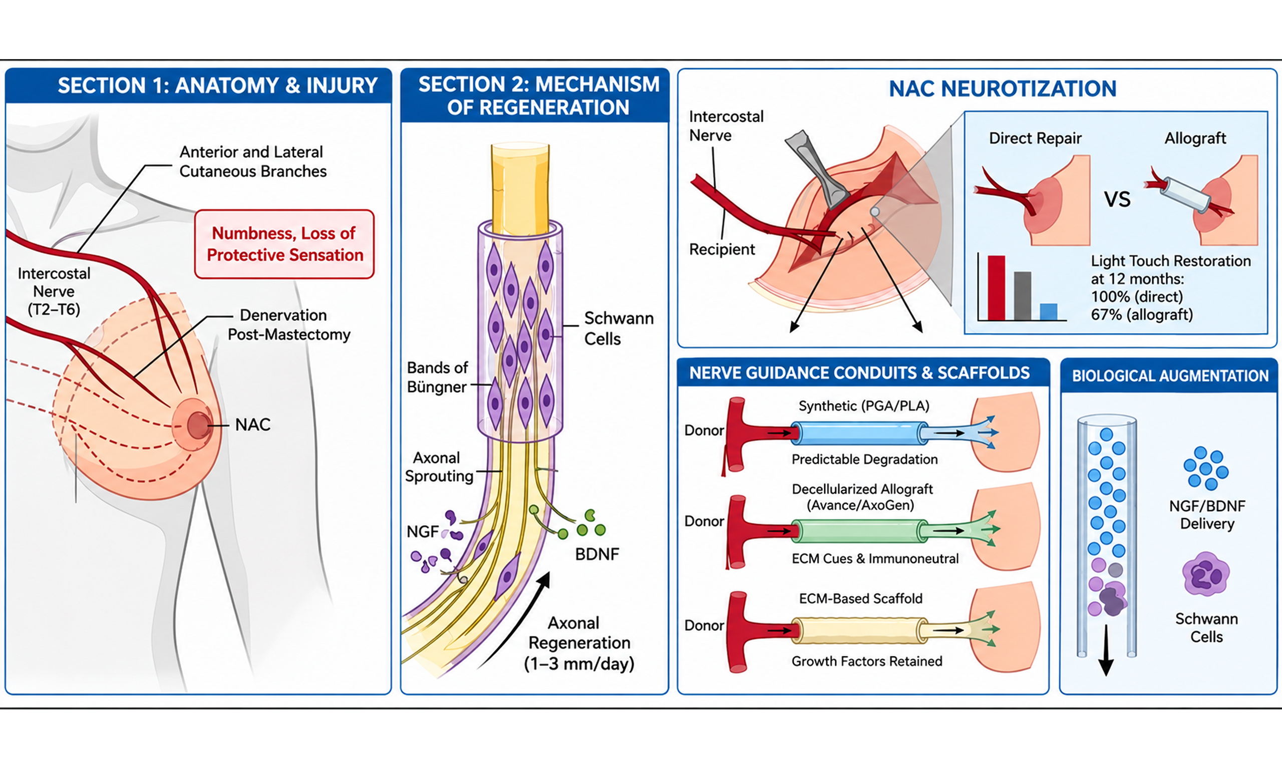
Sensory deficit is among the most prevalent long-term morbidities in the post-mastectomy reconstructed population, with direct implications for quality of life, body image, psychosexual function, and patient safety. Routine sacrifice of intercostal sensory nerves during mastectomy partially or completely denervates the breast skin and nipple-areola complex (NAC), producing numbness, paresthesia, and loss of protective sensation. Microsurgical neurotization, with or without nerve grafts, achieves clinically relevant sensory recovery in both autologous and implant-based reconstruction; meta-analytic evidence shows that neurotized flaps have superior tactile thresholds, faster recovery, and better protective sensation than non-neurotized flaps, although native sensation is not fully restored. Recovery is moderated by radiation history, regeneration distance, patient age, and surgical technique, and the field remains limited by donor-nerve availability, graft-related morbidity, cost, and the absence of standardized multimodal sensory assessment. Advances in regenerative medicine and biomaterials have introduced nerve guidance conduits, decellularized allograft scaffolds, extracellular matrix (ECM)-based conduits, neurotrophic factor delivery systems, and sutureless devices such as Nerve Tape (Biocircuit Technologies; Atlanta, GA), shifting emphasis from structural replacement toward biological restoration. This narrative review synthesizes clinical, translational, and mechanistic evidence on regenerative neurotization in breast reconstruction: it reviews peripheral nerve regeneration, appraises microsurgical neurotization and its comparative effectiveness, and evaluates regenerative adjuncts including biomaterial scaffolds, conduit-based repair, and molecular augmentation. A clinical translation framework is proposed, with sensory outcomes, methodological constraints, and future directions - smart biomaterials, artificial intelligence (AI)-assisted nerve mapping, and personalized regenerative reconstruction - identified as priorities for investigation.
Keywords
INTRODUCTION
Reconstructive surgery following mastectomy has historically prioritized volume and symmetry[1]; however, with improved survivorship, the focus has expanded to sensory restoration, which is a key determinant of quality of life, body image, and patient safety[2,3]. Sensory deficit is the most prevalent long-term morbidity in this population[4], arising from routine intercostal nerve sacrifice during mastectomy, which causes partial or complete denervation of the breast skin and nipple-areola complex (NAC)[5], manifesting as numbness, dysesthesia, loss of protective sensation, and risk of thermal injury[6].
Conventional neurotization restores sensation via donor-to-recipient nerve coaptation, improving protective and erogenous sensation with direct implications for psychosexual wellbeing[7]. Its limitations, which include the availability of donors, autograft-related morbidity, variable success rates, and the distance of regeneration[8], have also sparked interest in the development of alternative forms of regeneration. Allografts have reduced the donor-site morbidity but have also brought about problems related to cost-effectiveness and integration[9]. Nerve guidance conduits and bioengineered scaffolds have also been identified as being useful in the regeneration of nerves[10-13]. However, the literature on the effectiveness of nerve conduits is scattered across surgical, neuroscientific, and biomaterials literature, with varying methodologies[14]. This manuscript is a structured narrative review, employing a reproducible database search strategy with defined eligibility criteria while retaining interpretive rather than pooled synthesis. This approach enables critical evaluation across heterogeneous surgical, biomaterials, and neuroscience evidence that primary studies and meta-analyses cannot integrate in isolation, and is consistent with the narrative review article type as defined by the journal’s author guidelines[15].
BIOLOGICAL BASIS OF NERVE REGENERATION
Breast innervation anatomy and clinical relevance of denervation
Breast sensation is innervated by the anterior and lateral cutaneous branches of the intercostal nerves from T2 to T6, and the fourth intercostal nerve is the major NAC innervation[16-18]. The segmental denervation resulting from mastectomy cannot be compensated by single-nerve restoration alone; the NAC, given its functional and psychosexual significance, remains the primary neurotization target. The consequent numbness, thermal insensitivity, and loss of erogenous and protective sensation indicate the inadequacy of spontaneous reinnervation and the necessity of active surgical intervention.
Mechanisms of peripheral nerve regeneration
Following injury, Wallerian degeneration occurs distally while proximal sprouting regeneration occurs at 1-
Barriers to nerve regeneration in reconstructed breasts
In addition, the process of nerve regeneration within the reconstructed tissue is hindered by ischemia, disruption of the ECM by scarring, and the fact that the distances may be beyond the survival time of the endoneurial tubes[24-26], thus providing a rationale for active neurotization. Radiation also augments each of these barriers through accelerated fibrosis, vascular compromise, and decreased Schwann cell viability[27], which explains why successful coaptation alone does not ensure full restoration, leading logically to the biological augmentation of the repair microenvironment.
PRINCIPLES OF SURGICAL NEUROTIZATION
Surgical neurotization includes microsurgical coaptation of donor intercostal nerves to the recipient site within the flap or chest wall. This method is more efficient than the natural recovery process[28]. The anterior branch of the third intercostal nerve, which is identifiable at the third rib-sternal junction in 80% of all patients, is the primary target[29]. Direct tension-free coaptation remains the preferred technique. Where gap length precludes primary repair, processed nerve allografts provide a viable bridge with reduced donor-site morbidity compared to autografts[30]. Nerve autografts have historically been considered the gold standard for gap repair; however, their associated donor-site morbidity and the improving outcomes data for decellularized allografts have shifted practice preference in the breast reconstruction context toward allograft bridging as the primary non-direct option[30]. The timing of the procedure is an important prognostic factor. Index-stage neurotization provides the longest time window for nerve regeneration. In contrast, a delayed procedure results in a suboptimal sensory outcome because of the degeneration of the endoneurium during the time gap[31]. Patient-specific factors, graft length, history of irradiation, and wound healing capacity make the procedure more individualized. Therefore, it is important to have realistic preoperative discussions regarding the expected sensory outcome rather than the ideal outcome[32].
Indications and technical approach differ by reconstructive modality. In autologous reconstruction, direct intercostal nerve coaptation within the flap is the preferred strategy, supported by the largest body of comparative evidence. In implant-based and nipple-sparing mastectomy reconstruction, nerve preservation at the time of skin flap elevation combined with processed allograft bridging to the NAC represents the primary available strategy, though the supporting evidence is limited to small prospective series and feasibility data. In bilateral reconstruction, the authors suggest that neurotization of at least one breast may be considered, with bilateral repair pursued where anatomical and operative constraints permit; this represents the authors’ perspective rather than an established standard of care.
Mastectomy approach, nerve length, and oncologic considerations
The feasibility of tension-free direct coaptation - the technique associated with the most favorable NAC outcomes - is determined in large part by the length of recipient nerve preserved during mastectomy, which is in turn a function of the oncologic surgical technique. Conventional mastectomy transects the lateral cutaneous branches of the intercostal nerves at the chest wall, leaving stumps too short for primary repair and necessitating graft interposition. Several modifications increase usable nerve length. Deliberate preservation of the lateral intercostal nerve branches within the subcutaneous tissue of the lateral mastectomy skin flap, undertaken only where the branch can be spared without leaving glandular tissue behind, retains a longer recipient stump for coaptation[33,34]. A complementary strategy elongates the resected fourth intercostal cutaneous branch using the intercostal main nerve harvested from beneath the adjacent rib through the same mastectomy wound, generating an autograft of approximately 9 cm that reaches the nipple base without a separate donor incision and permits direct neurotization of the spared nipple and skin[5]. Nipple-sparing mastectomy is particularly amenable to these approaches because the preserved skin envelope and NAC provide defined distal targets, and immediate direct-to-implant reconstruction allows graft length to be tailored intraoperatively[33,35].
The governing oncologic principle is that nerve preservation must never compromise the completeness of the cancer operation: no glandular breast tissue is intentionally retained for the sake of preserving a nerve, and branches coursing through tissue that must be resected are transected and reconstructed by graft rather than spared[33]. On this basis, sensory-preserving technique does not alter the oncologic resection and therefore carries no established increase in local recurrence risk; this inference nonetheless rests on the same absence of long-term comparative recurrence data that characterizes the field as a whole, and prospective oncologic surveillance of neurotized cohorts remains an unmet need. Indication selection follows accordingly. The most straightforward candidates are patients undergoing risk-reducing (prophylactic) mastectomy and those undergoing nipple-sparing mastectomy for early-stage disease without an oncologic contraindication to nipple preservation, in whom favorable nerve anatomy can be exploited without deviation from the standard resection[33,35]. Patients with locally advanced disease, those requiring extensive skin or nipple resection, and those with anticipated post-mastectomy radiation - which independently degrades the regenerative microenvironment - are less suitable, and in this group the balance favors deferring or foregoing sensory-preserving maneuvers in favor of oncologic priorities.
The addition of neurotization introduces a distinct, if modest, complication profile that warrants disclosure during consent. Dissection of intercostal recipient or donor nerves toward the chest wall approaches the parietal pleura, and pleural violation is the most consequential potential event. In the largest comparative series to date, harvesting the intercostal main nerve was associated with pneumothorax in 5% of patients vs. 3% of controls and with a higher rate of pleural effusion (12% vs. 4%; P = 0.037), although the majority of affected patients were asymptomatic and managed conservatively[35]. Reported respiratory-function compromise after intercostal nerve harvest is otherwise minimal and largely subclinical[5]. Local morbidity includes the potential for painful neuroma and dysesthesia at the coaptation or donor site, though contemporary nipple-sparing series employing careful nerve handling report an absence of persistent dysesthesia or clinically evident neuroma[33]. Harvesting a long mixed intercostal segment additionally risks iatrogenic denervation of the rectus abdominis with resultant bulge, a consideration that has shifted preference toward selective sensory coaptation and interpositional allografting over extensive autograft harvest[34]. Overall, the incremental risk of sensory restoration is low but non-zero, and that technique selection should weigh donor-site and pleural risk against the anticipated sensory benefit for the individual patient.
METHODOLOGY
The search strategy and eligibility criteria are described below. This review retains interpretive rather than pooled synthesis, enabling critical evaluation across heterogeneous surgical, biomaterials, and neuroscience evidence that primary studies and meta-analyses cannot integrate in isolation.
Search strategy
Five databases were searched (PubMed, Scopus, Web of Science, ScienceDirect, and Embase) using terms including “breast reconstruction”, “neurotization”, “nerve regeneration”, “nerve guidance”, “regenerative biomaterials”, and “sensory restoration”; full search strings and yield by database are reported in Supplementary Table 1.
Eligibility criteria
Included studies reported clinical sensory restoration or neurotization outcomes in breast reconstruction, preclinical nerve regeneration mechanisms, or biomaterial-based nerve repair, published in English peer-reviewed journals. Excluded were cosmetic breast studies, mechanistically irrelevant case reports, and non-peer-reviewed material. A complete summary of included studies, reconstruction types, neurotization techniques, assessment methods, and key findings is provided in Supplementary Table 2.
EVIDENCE SYNTHESIS
Biological mechanisms of nerve regeneration in breast reconstruction
Peripheral nerve regeneration following breast reconstruction is a gradual and inherently incomplete process, the extent of which is determined by a hierarchy of biological, surgical, and patient-level variables. In the case of deep inferior epigastric perforator (DIEP) flap reconstruction, objective sensory improvement is reported to be partial, and the Semmes-Weinstein monofilament (SWM) score improves by a mean of -0.48 (P < 0.001) for DIEP specifically and -0.67 for all autologous modalities (P < 0.05)[36,37], although light touch and pinprick sensation only recover to 28% and 50% of contralateral breast values at 18 months. This disparity between improvement and restoration is the biological problem that regenerative adjuncts seek to address.
The most significant factor is the status of neurotization, with the average SWM of 4.24 ± 0.68 (mean ± SD) for the neurotized group compared to 5.06 ± 0.67 for the non-neurotized group; data from the pressure-specified sensory device (PSSD) confirmed the significance of the difference, with a calculated value of -13.36 (P = 0.0448)[38]. However, the significance of the difference is not just statistical but clinically relevant as well, as lower values of the SWM indicate the capacity for finer discriminatory sensation as well as greater protective sensibility[39]. Protective sensation over the majority of the breast surface is present in 55% of the neurotized patients compared to 7% of the non-neurotized patients (P < 0.01). Conversely, the absence of protective sensation is present in 27% of the neurotized patients compared to 64% of the non-neurotized patients[40]. Protective sensation over five segments of the breast was present in 75% of the neurotized DIEP flaps, 31% of the non-neurotized DIEP flaps, and 18% of the TRAM flaps, with absent somatosensory evoked potentials (SSEP) present in 0%, 23%, and 46% of the patients, respectively[41]. Overall, the evidence suggests that the effect of neurotization is not simply to augment sensation as a secondary effect[42]. Rather, the effect is fundamental to the chance of achieving meaningful sensory outcomes.
Recovery is also time-dependent in a manner that is highly relevant to surgical planning as well as counseling patients. Neurotized flaps demonstrate monthly SWM threshold reductions of 0.083 compared with 0.012 in non-neurotized flaps (P < 0.001) - a seven-fold difference in recovery rate[43]. Nevertheless, such enhanced speed is not sustained indefinitely. Sensory recovery reaches a plateau or slows substantially after 95 weeks at multiple breast locations (P < 0.05)[29]. The two-year mark therefore reflects a plateau rather than continued recovery. This effect is seen long before the full restoration of normal sensation is observed, which is not attained even in the highest-performing cohorts with a mean SWM of 2.71 ± 0.40 (mean ± SD) compared to the unoperated breast normal value[43].
Radiation history is a biologically significant and clinically underappreciated factor that compounds sensory recovery outcomes. Irradiated patients have been shown to have lower sensory scores compared to their non-irradiated counterparts (11.6 ± 6.7 vs. 16.3 ± 6.2; P = 0.012)[44]. Non-neurotized breasts are more than twice as likely to have been irradiated as neurotized breasts, 47.4% as opposed to 20.7%[27]. This indicates that irradiation can serve as a negative prognostic modifier as well as a preventative measure for neurotization[45], which is a compounding factor that requires specific attention.
Microsurgical neurotization techniques for sensory restoration
Microsurgical neurotization of the breast is the first technique for the restoration of breast sensation, demonstrating superiority over non-neurotized controls. The standard technique involves end-to-end anastomosis to the anterior branch of the third intercostal nerve, identifiable at the third rib-sternum junction in 80% of patients, accomplished with 9-0 nylon suture or a 40 mm conduit[29]. Where direct coaptation is not possible, allografts of 50-70 mm length and 1-2 mm diameter bridge the gap, with technical success in 85% of planned cases[46].
Evidence for technique superiority is consistent across flap modalities. In transverse rectus abdominis myocutaneous (TRAM) flap series, 45% sensation recovery relative to the contralateral breast was achieved in neurotized patients vs. 26% in controls, with 12 of 13 neurotized patients demonstrating superficial tactile sensation vs. 1 of 13 non-neurotized[47]. PSSD-measured static touch was imperceptible at the flap center in 14.3% of innervated vs. 42.1% of non-innervated flaps at 24 months (P = 0.015), with heat pain imperceptible in 10.3% vs. 42.1% respectively (P = 0.004)[27]. A meta-analysis of 352 DIEP flaps demonstrated a pooled SWM benefit of -0.86 (P < 0.05) independent of age, chemotherapy, and radiotherapy[37] - a finding that is notable for suggesting that neurotization benefit persists even in biologically compromised patients[48].
For NAC-specific recovery, direct coaptation demonstrates superiority over allograft bridging: at 12 months, 100% of directly coapted patients showed restored NAC light touch vs. 67% in the allograft group and 17.5% of non-neurotized controls (P < 0.05), with subjective NAC sensation restored in 95%, 75%, and 4% respectively[49]. Comparable superiority of direct coaptation has been reported in gender-affirming mastectomy cohorts[50]. This difference extends beyond tactile function: the NAC carries erogenous and psychosexual significance, and its reinnervation has been associated with improved body image and sexual wellbeing; this evidence derives substantially from nipple-preservation and gender-affirming mastectomy cohorts and is extrapolated to the post-mastectomy setting[7,51] (Section “Erogenous sensation and cortical reorganization”).
Nerve Tape (Biocircuit Technologies; Atlanta, GA), a recently introduced sutureless coaptation device, reduces repair time to 1.79 ± 1.71 min vs. 5.20 ± 2.20 min for conventional microsuture (P = 0.0002), with 92% of repairs rated clinically acceptable vs. 69% for microsuture, and 85% of trainee surgeons completing repair within 2 min[52]. These figures have significant implications not only for operative efficiency but for broadening access to neurotization - a technically demanding procedure whose adoption has been limited by microsurgical expertise requirements.
Biomaterials and tissue-engineering approaches for guided nerve regeneration
Biomaterial scaffolds for nerve repair range from structurally passive conduits to biologically active composites, and their systematic evaluation in breast reconstruction remains at an earlier stage than in peripheral nerve surgery more broadly. The key translational question is not whether conduits can bridge nerve gaps - that is now well established - but whether specific material properties can actively promote the quality and speed of axonal regeneration in the more clinically challenging environment of reconstructed, often irradiated, tissue where preclinical gap-repair models offer limited predictive validity.
Synthetic conduits composed of polyglycolic acid (PGA) and polylactic acid (PLA) provide predictable degradation profiles. Comparative threshold data for bridged vs. direct repair are available from a nerve allograft series rather than a synthetic conduit series: in a retrospective comparison of 53 DIEP flaps, sensory thresholds were 78.28 g/mm2 with nerve allograft (35 flaps) vs. 64.58 g/mm2 with direct coaptation (18 flaps), a difference that was not statistically significant (P = 0.680)[49]. A non-significant P-value indicates only that no difference was detected with the available sample and does not, by itself, establish statistical equivalence; a formal equivalence or non-inferiority analysis with a predefined margin and corresponding confidence intervals would be required to support that conclusion, and this has not been reported for allograft or PGA/PLA conduit repair in breast neurotization. Threshold parity, even if confirmed, should in any case not be interpreted as equivalence in recovery trajectory or functional outcome range, nor extrapolated to synthetic conduits, which offer no biological interaction with the regenerating axon, providing geometry without guidance.
Decellularized nerve allografts (e.g., Avance, AxoGen; Alachua, FL) preserve native endoneurial architecture and ECM molecular cues while eliminating immunogenicity. NAC neurotization with Avance showed moving sensibility at 12 months in 69%-73% of breasts (92% in the upper quadrant) and two-point discrimination preserved in 87%[33], supporting decellularized allografts as the preferred non-autologous bridging material - particularly in implant-based reconstruction where allograft bridging is the primary available strategy[53].
ECM-based conduits (small intestinal submucosa, urinary bladder matrix) offer bioactive scaffolds with retained growth factors and adhesion molecules, though clinical translation in breast reconstruction remains limited and comparative data against allografts are lacking[54]. Collagen conduits occupy an intermediate bioactivity position and retain utility for short-gap repairs[55].
Coaptation number is an underexplored variable: double neurorrhaphy with conduits in muscle-sparing TRAM procedures outperformed single neurorrhaphy over 24-43 months follow-up[47], suggesting that expanding the total area of guided reinnervation may matter more than optimizing a single repair - an important consideration in patients with larger resection volumes.
Biological augmentation strategies and translational context
Despite optimized neurotization, complete sensory restoration remains unachieved: neurotized TRAM patients reach ~45% of contralateral sensation, SWM scores [4.24 ± 0.68 (mean ± SD)] fall short of the unoperated breast norm (2.66 ± 0.65)[38], and complete sensory absence persists in a minority of neurotized patients vs. a substantially larger proportion of non-neurotized controls. This gap defines the rationale for biological augmentation strategies targeting the regenerative microenvironment.
Neurotrophic factor delivery represents the most biologically grounded augmentation approach. NGF and BDNF sustain Schwann cell activity and axonal regenerative momentum; their depletion following endoneurial tube degeneration is a primary cause of failed long-distance regeneration. Localized delivery of microspheres, hydrogel depots, or factors within the conduit walls aims to prolong the permissive regeneration period beyond the capacity of the intrinsic axonal growth rate (1-3 mm/day)[10,11]. Neurotrophic factor delivery currently remains at the preclinical stage of translational development [technology readiness level (TRL) 3-4]. While the biological rationale is well-established in animal models of peripheral nerve repair, no clinical series in breast reconstruction has been reported. Translation is contingent on resolving delivery kinetics, scaffold integration, and regulatory pathway requirements prior to first in-human application.
Beyond the neurotrophins themselves, attention has turned to which specific agents are most promising for clinical translation and to whether any systemically administered drug already in human use can accelerate reinnervation. Among growth factors, NGF, BDNF, glial cell line-derived neurotrophic factor (GDNF), and vascular endothelial growth factor have the strongest preclinical support, with GDNF notable for its potency in promoting sensory neuron survival; the principal translational obstacle common to all is the engineering of sustained, spatially controlled release rather than efficacy in animal models, since bolus delivery is rapidly cleared and supraphysiologic concentrations can paradoxically trap regenerating axons at the release site. The most clinically advanced pharmacologic candidate is tacrolimus (FK506), an immunosuppressant in widespread human use whose neuroregenerative properties are independent of, and occur at lower doses than, its immunosuppressive action. In rodent nerve repair, systemic FK506 accelerates axonal outgrowth and increases myelinated fiber density, raising the sensory regeneration rate from approximately 3.8 to
A further implication follows from the mechanism itself. The neurotrophic action of FK506 is calcineurin-independent, mediated not by FK506-binding protein 12 (FKBP-12) (the effector of immunosuppression) but by the immunophilin FK506-binding protein 52 (FKBP-52), such that its regenerative and immunosuppressive activities are pharmacologically dissociable[58]. This dissociation gave rise to non-immunosuppressive FKBP ligands (e.g., GPI-1046 and V-10367) that retained axonal-regenerative activity without immunosuppression in preclinical models[59]. Although clinical translation of these analogs subsequently stalled, the concept is of particular relevance to the oncologic breast population, in whom systemic immunosuppression is not merely a generic toxicity but a theoretical liability for immunosurveillance of residual or micrometastatic disease. Two features therefore make tacrolimus, or a non-immunosuppressive congener, suited to this setting: the neurotrophic effect occurs at doses below the immunosuppressive threshold, and localized delivery further restricts systemic exposure to negligible levels, together decoupling regenerative benefit from the oncologic concern that would otherwise preclude its use. Unlike novel neurotrophic biologics, moreover, tacrolimus is already in wide human use, so a drug-eluting graft or conduit constitutes a repurposing pathway rather than a new molecular entity - plausibly the shortest translational route among the augmentation strategies reviewed here. The principal caveats are that existing regeneration data are predominantly motor or mixed-nerve and rodent, with no sensory-specific or breast-reconstruction evidence, and that any oncologic-safety claim for local delivery remains to be demonstrated rather than assumed.
Schwann cell seeding in conduits simulates the native nerve biological guidance cues. Although autologous cells from non-critical nerves of donors circumvent immunogenicity[21], the clinical application is hampered by the intricacy and expense of good manufacturing practice (GMP)-grade ex vivo expansion protocols. Schwann cell-based strategies therefore currently remain at TRL 2-3, with translation relying on advances in cell expansion technology and regulatory frameworks for autologous cell therapy products. Age was the strongest predictor when examined as a standalone factor (independent predictor: P = 0.004 moving touch, P = 0.008 static touch); however, radiotherapy was another strong factor[27,60]. These patients present the highest unmet clinical need with the lowest intrinsic regenerative capacity and thus require validation of the augmentation strategy in compromised biological environments as opposed to healthy younger cohorts.
Modality-specific sensory recovery
A recurring interpretive limitation of the breast neurotization literature is the treatment of “sensation” as a single quantity, when the reinnervated breast in fact reacquires several functionally and anatomically distinct submodalities at different rates and to different degrees. Cutaneous sensation is subserved by mechanoreceptors (detecting light touch, pressure, vibration, and stretch), thermoreceptors (warm and cool detection), and nociceptors (signaling actual or potential tissue damage as pain), with proprioceptive contribution from deeper structures; protective sensation - the clinically pivotal endpoint that guards against thermal and mechanical injury - depends chiefly on the nociceptive and thermal channels rather than on the fine touch that monofilament testing captures. The distinction is relevant because the instrument used in roughly 94% of studies, the SWM, interrogates only the low-threshold mechanoreceptive channel and is therefore blind to precisely the modalities most relevant to patient safety. The limited studies that have applied broader quantitative sensory testing show that recovery is genuinely modality-specific: in a double-blind trial, innervated flaps regained not only tactile thresholds but also thermal and heat-pain perception, with static touch imperceptible at the flap center in 14.3% of innervated vs. 42.1% of non-innervated breasts and heat pain imperceptible in 10.3% vs. 42.1%, respectively[27]. Comprehensive quantitative sensory testing in the closely related setting of gender-affirming mastectomy with targeted reinnervation has demonstrated simultaneous improvement across mechanical detection, two-point discrimination, vibration, pinprick, temperature detection, heat-pain, and pressure-pain thresholds, confirming that a single mechanoreceptive threshold substantially underrepresents the sensory result[61]. Two practical implications follow. Outcome batteries should sample mechanoreceptive, thermal, and nociceptive channels rather than tactile threshold alone, and counseling should frame recovery of protective (thermal and nociceptive) sensation - not fine touch - as the primary safety-relevant goal.
Erogenous sensation and cortical reorganization
Loss of erogenous sensation is among the least measured consequences of mastectomy, and its restoration is a distinct objective from protective or discriminative sensation because the NAC carries an erogenous function separate from that of the surrounding breast skin[7]. Direct evidence in oncologic reconstruction remains sparse, but the gender-affirming mastectomy literature - which uses the same intercostal donor nerves and NAC targets - now provides the most relevant data. In a prospective matched cohort undergoing gender-affirming mastectomy, targeted NAC reinnervation improved patient-reported erogenous sensation alongside objective sensory measures relative to non-reinnervated controls[61], and a systematic review reported that approximately 88% of reinnervated patients regained erogenous sensation by twelve months, with correspondingly higher satisfaction on patient-reported measures[62]. These findings support the inference, still requiring direct confirmation in the oncologic setting, that reestablishing afferent input to the NAC is a prerequisite for erogenous recovery and that its inclusion as a coaptation target carries psychosexual value beyond tactile function.
The relationship between restored peripheral input and subjective erogenous experience is not, however, purely peripheral; the reconstructive-genital literature offers parallels in cortical reorganization. Deafferentation produces measurable reorganization of the primary somatosensory cortex, and the perceptual quality of sensation arising from reinnervated tissue depends on how the central representation accommodates the returning input[22,23]. Genital reconstruction provides the clearest clinical analog: sensory recovery after genitoplasty is reportedly faster and more complete than in most other peripheral nerve scenarios, and erogenous sensation - including orgasmic capacity - is frequently reported even where quantitative afferent recovery is modest[63]. This apparent dissociation between measured input and erogenous experience has been attributed to cortical plasticity, in the hypothesis that the cortex can interpret non-native afferent input as erogenous - that, functionally, the relevant cortex “feels what it expects to feel” - a mechanism proposed to underlie erogenous recovery in phalloplasty[64]. That genital and, on some accounts, nipple sensation occupy discrete cortical territory further suggests that erogenous reinnervation engages central circuitry not fully captured by peripheral threshold testing[65]. For the reconstructed breast, this implies that erogenous outcomes may depend jointly on the quality of peripheral coaptation and on central adaptation, and that patient-reported erogenous measures capture a dimension of recovery that no cutaneous instrument captures, supporting their use as primary endpoints.
Clinical outcomes and evidence synthesis
The evidence across flap modalities, patient demographics, and outcome instruments converges on a consistent finding: surgical neurotization produces clinically meaningful and reproducible sensory benefits that spontaneous reinnervation does not replicate. The magnitude of this benefit, a pooled SWM improvement of -0.86 in DIEP subgroup analysis, a seven-fold difference in monthly recovery rate, and a doubling of protective sensation prevalence, is notable for its consistency across cohorts that differ substantially in age, radiation history, and chemotherapy exposure[37,40,43]. These findings carry direct quality-of-life implications: protective sensation, NAC reinnervation, and erogenous recovery each represent functional endpoints whose clinical significance extends beyond threshold measurements alone[51]. Direct coaptation demonstrates superiority over allograft bridging for NAC-specific recovery, with 100% vs. 67% light touch restoration at 12 months (P < 0.05), though both strategies substantially outperform non-neurotized controls[49] [Table 1].
Key evidence summary
| Study | Design | Key finding | n | LOE | Risk of bias | Other limitations |
| Aristizábal et al. (2025)[37] | SR/MA | SWM MD -0.86 favor neurotized (P < 0.05) | 352 (DIEP subgroup; 495 breasts overall) | I | Risk of bias in constituent primary studies not systematically appraised (no AMSTAR-2/ROBIS tool applied) - a review-level appraisal gap, not participant-level selection bias | Heterogeneity in coaptation technique; no standardized follow-up interval |
| Tajziehchis et al. (2024)[38] | SR/MA | SWM MD -0.88; PSSD MD -13.36 | 12 studies (367 neurotized, 295 non-neurotized flaps) | I | Risk of bias in constituent primary studies not systematically appraised (no AMSTAR-2/ROBIS tool applied) | Mixed flap types; variable outcome instruments |
| Bubberman et al. (2024)[27] | Preliminary RCT | Static touch imperceptible: 14.3% vs. 42.1% | 41 | II | None identified - randomization and double-blind assessment substantially mitigate selection and detection bias | Small n = 41; single center; preliminary designation (underpowered) |
| Bijkerk et al. (2020)[46] | Prospective cohort | Innervated flaps: significantly better sensation vs. non-innervated (t-test, P < 0.05) | 15 | II | Selection bias: non-random allocation of side to neurotization. Detection bias: no blinding of outcome assessors to neurotization status | n = 15; within-patient design limits generalizability across patients |
| Momeni et al. (2021)[40] | Prospective cohort | Protective sensation: 55% vs. 7% (P < 0.01); absent protective sensation: 27% vs. 64% | 59 | III | Selection bias: non-randomized allocation; referral pattern toward high-volume, neurotization-experienced centers (registry-based) | n = 59; registry design limits causal inference |
| Zhang et al. (2025)[49] | Retrospective | NAC light touch @12 months: 100% direct vs. 67% allograft | 53 | III | Selection bias: retrospective, non-randomized allocation to direct coaptation vs. allograft | n = 53; single-surgeon series limits generalizability |
Complete sensory restoration remains unachieved across all reported cohorts, with neurotized SWM scores consistently falling short of the unoperated breast norm[43]; the goal of neurotization is therefore meaningful improvement within a biologically constrained ceiling, not equivalent to native sensation. The current evidence base, predominantly level of evidence (LOE) II-III and including two small randomized trials [a completed randomized controlled trial (RCT) in free TRAM flap reconstruction[66] and the preliminary, interim results of an ongoing double-blind RCT in DIEP flap reconstruction[27]; Section “Limitations and the case for outcome standardization”], supports a Grade B evidence-informed practice recommendation for neurotization in eligible autologous reconstruction, consistent with Oxford Centre for Evidence-Based Medicine criteria. The consistency of benefit across a heterogeneous reconstructive literature represents a level of concordance that is uncommon in reconstructive surgery; guideline-level designation awaits the results of well-powered multicenter randomized trials.
DISCUSSION
Surgical neurotization leads to clinically important, reproducible sensory improvement, and the biological potential of the regenerative adjuncts is real in terms of bridging the gap to the desired restoration. However, the evidence also shows a field that is methodologically fragmented, lacks well-developed comparative evaluation of biomaterials, and still suffers from a disconnect between the emphasis placed on regenerative strategies in the literature, including the title of a review, and the quality of the controlled evidence. This disconnect is the methodological challenge facing the field.
Three themes warrant emphasis. First, the benefits of neurotization are consistent across flap types, patient demographics, and outcome measures, including a seven-fold difference in monthly recovery rate, a doubling of protective sensation prevalence, and a pooled SWM benefit irrespective of age, chemotherapy, or radiotherapy[37]. These findings support a Grade B evidence-informed practice recommendation for neurotization in eligible autologous reconstruction, consistent with Oxford Centre for Evidence-Based Medicine criteria. The persistence of benefit in patients least expected to respond, including those with prior radiotherapy or advanced age, strengthens the surgical rationale precisely in the population where decision-making is most complex. Second, the evidence supporting biomaterials and augmentation is substantially weaker than that supporting neurotization. Conduit comparison, ECM scaffold optimization, and neurotrophic factor translation are all outstanding, warranting greater prominence than they are afforded in the literature, including dedicated translational research to substantiate the pre-eminence of these approaches. Third, the variety of outcome measures, including SWM, PSSD, thermal testing, evoked potentials (EPs), and patient-reported outcomes (PROs), makes direct comparison between studies difficult and likely results in a systemic underestimation of the benefits of neurotization, particularly in those studies where less sensitive outcome measures are utilized. This is addressed in Section “Limitations and the case for outcome standardization”, where the importance of standardization is highlighted.
CURRENT LIMITATIONS, CONTROVERSIES, AND FUTURE DIRECTIONS
Limitations and the case for outcome standardization
A structured appraisal of study quality reveals consistent methodological limitations across the included evidence base. The majority of primary studies are single-center prospective cohorts or retrospective series (LOE II-III) performed at high-volume microsurgical centers, introducing probable selection bias toward technically favorable cases, younger patients, and lower rates of prior irradiation. The two available systematic reviews and meta-analyses, while providing the highest available level of evidence, are themselves constrained by the heterogeneity of their constituent studies, differing in flap type, coaptation technique, outcome instrument, and follow-up duration. Randomized evidence on patient sensory outcomes remains limited to two small trials of different eras and designs: an earlier RCT of innervated vs. non-innervated free TRAM flaps with blinded sensory assessment (Temple et al., 2006; n = 27)[66], and the preliminary, interim results of an ongoing double-blind RCT in DIEP flaps (Bubberman et al., 2024; n = 41)[27], which was underpowered for definitive conclusions. No included study employed evidence synthesis using the Grading of Recommendations Assessment, Development and Evaluation (GRADE) framework or formal risk-of-bias assessment using validated instruments such as the Risk Of Bias In Non-randomized Studies of Interventions (ROBINS-I) or the Revised Cochrane risk-of-bias tool for randomized trials (RoB 2). These limitations are reflected in the evidence-informed rather than guideline-level strength of recommendations presented in this review. It is also worth noting that the structured, reproducible data-curation approach adopted here satisfies many of the methodological criteria for a scoping review; although the present work is framed as a narrative synthesis consistent with the journal’s article types, formal scoping- or systematic-review methodology represents a natural and higher-evidence progression for future iterations of this question.
The most critical limitation is the lack of a standardized multimodal assessment tool set. Although SWM, utilized in ~94% of all studies, has a proven ceiling effect of two-point discrimination pre-operatively in 10%-11% of patients[42,66], whereas the more sensitive PSSD is utilized in merely 12.5% of all studies. This review proposes a minimum standard of measurement: a threshold measurement (SWM), a discrimination measurement (PSSD), and the BREAST-Q: Breast Sensation Module as a reconstructive-specific PRO measure. Uniform implementation across prospective cohorts and RCTs would enable inter-study comparison, subgroup analysis by flap type and radiation status, and patient-centered evidence for regulatory decisions.
Additional limitations include small, demographically narrow sample sizes - some cohorts include as few as 16 participants[33,67] - which limit generalizability to older, higher-BMI, or more complex reconstructive populations. Follow-up durations rarely exceed 24 months despite evidence of continued sensory change beyond this point. Substantial variability in flap type, graft material, coaptation number, and radiation exposure (5.7%-64% across comparative cohorts) further constrains inter-study comparability. Cost and accessibility constraints on allograft material and microsurgical expertise create inequities in access to neurotization that are not adequately captured in the current literature, and protracted regulatory pathways for novel biomaterials continue to delay clinical translation of the most promising regenerative strategies.
Future directions
The following represent the highest-priority directions based on identified evidence gaps.
Smart biomaterials and electroactive conduits
Electroactive conduits incorporating piezoelectric or conductive polymers enhance Schwann cell alignment and axonal elongation in preclinical models. Combining these with timed neurotrophic factor release represents the next engineering objective, with particular urgency for irradiated patients whose static microenvironment least supports regeneration.
Localized neurotrophic delivery
Controlled NGF/BDNF release via microspheres or hydrogels at the repair site could extend the permissive regeneration window and compress the current 12-24-month assessment timeline, reducing patient burden.
Artificial intelligence (AI)-assisted nerve mapping
Standard anatomical landmarks identify the target nerve in only ~80% of patients[29]. AI-based image segmentation and analysis have shown high diagnostic accuracy for peripheral nerve identification on ultrasound and magnetic resonance imaging (MRI) in adjacent surgical fields, and are increasingly proposed as adjuncts for intraoperative anatomical localization[68]. Real-time AI-enhanced imaging integrating MRI tractography with intraoperative ultrasound or near-infrared fluorescence could reduce anatomical failure rates; this application remains untested in breast neurotization specifically and is extrapolated from feasibility data in adjacent surgical fields[68].
Robotic microsurgery
Tremor filtering and motion scaling could help improve anastomotic precision in fine-caliber nerve repair, thereby increasing neurotization access to revision cases and currently excluded patient cohorts on technical grounds.
Three-dimensional (3D) bioprinted personalized nerve guides
Patient-specific scaffolds seeded with autologous Schwann cells, geometrically and biologically matched to patient anatomy and radiation history, represent a regenerative endpoint. 3D-printed nerve guide conduits fabricated to patient-specific geometry have demonstrated feasibility and effective axonal regeneration in preclinical models[69], though seeding with autologous Schwann cells and application to the breast neurotization setting have not been reported. In the authors’ view, clinical translation may be achievable within approximately the next decade; this estimate is speculative and contingent on future preclinical validation, clinical testing, and regulatory progress.
The intercostobrachial nerve: an underrecognized sensory morbidity after axillary dissection
The neurotization literature is almost entirely concerned with restoring sensation to the breast skin and NAC, yet a substantial component of post-treatment sensory morbidity originates outside this territory, in the distribution of the intercostobrachial nerve (ICBN). The ICBN supplies sensation to the axilla and medial upper arm and is frequently divided during axillary lymph node dissection, producing numbness, paresthesia, and dysesthesia of the upper inner arm; anecdotally, and in some patient-reported series, this axillary and arm dysesthesia is at least as bothersome as breast or nipple numbness, in part because it is provoked by everyday arm movement and clothing contact. Despite its prevalence, this morbidity is rarely measured on the same instruments used for breast sensation, and direct patient-reported comparisons between ICBN-territory symptoms and breast or NAC numbness are essentially absent from the reconstructive literature - a gap that likely leads the field to underestimate the total sensory burden of breast cancer surgery.
Evidence on prevention centers on surgical handling of the nerve. A systematic review and meta-analysis of ICBN preservation vs. division during axillary dissection found that preservation reduces the incidence of anesthesia and hypesthesia within the nerve’s territory, although its effects on pain and paresthesia were less consistent[70]. Randomized data are mixed on objective sensory endpoints but suggest a quality-of-life benefit: a double-blind trial reported that preservation, while not uniformly improving measured sensation, improved health-related quality of life and upper-limb physical function in the early postoperative period[71]. Refinements in dissection technique, including energy-device rather than sharp division of surrounding tissue, have been explored as means of reducing nerve injury, and where the nerve is unavoidably transected the same coaptation and grafting principles applied to breast neurotization are, in principle, available for the ICBN, though this has not been systematically studied. Two priorities follow: PRO instruments for breast reconstruction should incorporate the axillary and arm sensory domain so that ICBN morbidity can be weighed against breast and NAC outcomes, and ICBN preservation should be regarded as a low-cost, quality-of-life-relevant adjunct to be pursued whenever oncologic clearance permits.
CLINICAL TRANSLATION FRAMEWORK
Table 2 consolidates indications, patient selection, technique preferences, and research priorities based on available evidence.
Clinical translation framework for regenerative neurotization
| Domain | Recommendation | Evidence base | Translational stage |
| Neurotrophic delivery | NGF/BDNF depot at repair site[10,11,56,57] | Preclinical only | TRL 3-4/Preclinical |
| Schwann cell seeding | Autologous cells in conduit[21] | Preclinical; GMP barriers | TRL 2-3/Preclinical |
| Smart conduits | Electroactive/piezoelectric[55] | Preclinical models | TRL 3/Preclinical |
| Decellularized allograft | Avance for gap bridging[33,53] | Clinical series | TRL 7-8/Clinical |
| Nerve tape | Sutureless coaptation[52] | Intraoperative RCT (repair quality/time, not patient sensory outcome) | TRL 6-7/Early clinical (device-handling feasibility; sensory outcomes not yet reported) |
| Synthetic conduits | PGA/PLA for short gaps | Non-inferiority not formally demonstrated (see text) | TRL 6-7/Early clinical |
For the post-mastectomy patient, the goals of reconstruction have expanded beyond volumetric restoration to include factors like sensory identity. Current evidence supports neurotization as the preferred approach in eligible autologous reconstruction, with a strength of recommendation commensurate with the available Level II-III evidence and the consistency of benefit observed across heterogeneous cohorts. Preoperative counseling should distinguish between achievable sensory improvement and the unattained benchmark of native sensation, a distinction that remains the honest basis of informed consent until randomized trial data mature.
CONCLUSION
Regenerative neurotization has moved from an experimental adjunct to evidence-based practice. Neurotized flaps have been shown to result in faster recovery, greater protective sensation, and better NAC reinnervation regardless of flap type, population, and assessment tool used. Sensory innervation remains unrestored, and biological barriers such as irradiation and age remain unsettled with surgical technique alone. The regenerative approaches described in this review - biomaterials, bioengineered scaffolds, neurotrophic factors, Schwann cells, and smart biomaterials - are a biologically grounded response to these challenges. For these regenerative approaches to be successfully translated into practice, more rigorous comparative data than currently available in the literature must be forthcoming. Standardization of sensory outcome measures in prospective cohorts using standardized tools such as SWM, PSSD, and BREAST-Q Breast Sensation Module as a minimum requirement, well-powered multicenter RCTs, and the incorporation of PROs as primary rather than secondary endpoints are the advances in study design and methodology without which this field cannot progress from promising evidence to conclusive, guideline-level practice change.
DECLARATIONS
Acknowledgments
The Graphical Abstract was created with BioRender.com.
Authors’ contributions
Study conception and design: Nadeem-Tariq A, Baynosa R
Literature search and data curation: Nadeem-Tariq A, Childers E
Manuscript drafting: Nadeem-Tariq A, Childers E
Critical revision for important intellectual content: Nadeem-Tariq A, Childers E, Baynosa R
All authors approved the final version of the manuscript for publication.
Availability of data and materials
Not applicable.
AI and AI-assisted tools statement
Not applicable.
Financial support and sponsorship
None.
Conflicts of interest
All authors declared that there are no conflicts of interest.
Ethical approval and consent to participate
Not applicable.
Consent for publication
Not applicable.
Copyright
© The Author(s) 2026.
Supplementary Materials
REFERENCES
1. Chae MP, Hunter-Smith DJ, Spychal RT, Rozen WM. 3D volumetric analysis for planning breast reconstructive surgery. Breast Cancer Res Treat. 2014;146:457-60.
2. Abdul-Al M, Zaernia A, Sefat F. Biomaterials for breast reconstruction: Promises, advances, and challenges. J Tissue Eng Regen Med. 2020;14:1549-69.
3. Inocenti A, Santos MAD, Loyola EACD, Magalhães PAPD, Panobianco MS. Impact of the effects of the reconstructive surgery in the life of women with breast cancer. Texto Contexto - Enferm 2016;25:e4520014.
4. Lovelace DL, McDaniel LR, Golden D. Long-term effects of breast cancer surgery, treatment, and survivor care. J Midwifery Womens Health. 2019;64:713-24.
5. Chang TN, Lu JC, Sung CW, et al. Elongation of intercostal nerve cutaneous branches for breast and nipple neurotization during breast reconstruction after mastectomy for breast cancer: case-control study. Br J Surg. 2024;111:znae005.
6. Joshua AM, Misri Z. Peripheral nerve disorders. In: Joshua AM, Editor. Physiotherapy for adult neurological conditions. Singapore: Springer Nature; 2022. pp. 621-729.
7. Deptula P, Nguyen D. Neurotization of the nipple-areola complex: superior nipple sensation in gender-affirming mastectomy and autologous breast reconstruction. Ann Breast Surg. 2021;5:11.
8. Bamba R, Loewenstein SN, Adkinson JM. Donor site morbidity after sural nerve grafting: A systematic review. J Plast Reconstr Aesthet Surg. 2021;74:3055-60.
9. Mahajan N, Soker S, Murphy SV. Regenerative medicine approaches for skin wound healing: from allografts to engineered skin substitutes. Curr Transpl Rep. 2024;11:207-21.
10. Panagopoulos GN, Megaloikonomos PD, Mavrogenis AF. The present and future for peripheral nerve regeneration. Orthopedics. 2017;40:e141-56.
11. Kong L, Gao X, Qian Y, Sun W, You Z, Fan C. Biomechanical microenvironment in peripheral nerve regeneration: from pathophysiological understanding to tissue engineering development. Theranostics. 2022;12:4993-5014.
13. Bell JH, Haycock JW. Next generation nerve guides: materials, fabrication, growth factors, and cell delivery. Tissue Eng Part B Rev. 2012;18:116-28.
14. Boecker A, Daeschler SC, Kneser U, Harhaus L. Relevance and recent developments of chitosan in peripheral nerve surgery. Front Cell Neurosci. 2019;13:104.
15. Barry ES, Merkebu J, Varpio L. Understanding state-of-the-art literature reviews. J Grad Med Educ. 2022;14:659-62.
16. Hamdi M, Würinger E, Schlenz I, Kuzbari R. Anatomy of the breast: a clinical application. In: Hamdi M, Editor. Vertical scar mammaplasty. Berlin: Springer Berlin Heidelberg; 2018. pp. 3-12.
18. Gupta M, Goyal N. Applied anatomy of breast cancer. In: Sharma SC, Mazumdar A, Kaushik R, Editors. Breast cancer. Singapore: Springer Nature; 2022. pp. 23-35.
19. Stoll G, Müller HW. Nerve injury, axonal degeneration and neural regeneration: basic insights. Brain Pathol. 1999;9:313-25.
20. Bradke F. Mechanisms of axon growth and regeneration: moving between development and disease. J Neurosci. 2022;42:8393-405.
21. Schuh CMAP, Sandoval-Castellanos AM, De Gregorio C, Contreras-Kallens P, Haycock JW. The role of Schwann cells in peripheral nerve function, injury, and repair. In: Gimble JM, Marolt Presen D, Oreffo ROC, Wolbank S, Redl H, Editors. Cell engineering and regeneration. Cham: Springer International Publishing; 2020. pp. 215-36.
22. Bubberman JM, Van Rooij JAF, Van der Hulst RRWJ, Tuinder SMH. Sensory recovery and the role of innervated flaps in autologous breast reconstruction-a narrative review. Gland Surg. 2023;12:1094-109.
23. Tataranu LG, Rizea RE. Neuroplasticity and nervous system recovery: cellular mechanisms, therapeutic advances, and future prospects. Brain Sci. 2025;15:400.
24. Rafique M, Ali O, Shafiq M, et al. Insight on oxygen-supplying biomaterials used to enhance cell survival, retention, and engraftment for tissue repair. Biomedicines. 2023;11:1592.
25. López-Cebral R, Silva-Correia J, Reis RL, Silva TH, Oliveira JM. Peripheral nerve injury: current challenges, conventional treatment approaches, and new trends in biomaterials-based regenerative strategies. ACS Biomater Sci Eng. 2017;3:3098-122.
26. Mahar M, Cavalli V. Intrinsic mechanisms of neuronal axon regeneration. Nat Rev Neurosci. 2018;19:323-37.
27. Bubberman JM, Brandts L, van Kuijk SMJ, van der Hulst RRWJ, Tuinder SMH. The efficacy of sensory nerve coaptation in DIEP flap breast reconstruction - Preliminary results of a double-blind randomized controlled trial. Breast. 2024;74:103691.
28. Pereira CT, Hill EE, Stasyuk A, et al. Molecular basis of surgical coaptation techniques in peripheral nerve injuries. J Clin Med. 2023;12:1555.
29. Spiegel AJ, Menn ZK, Eldor L, Kaufman Y, Dellon AL. Breast reinnervation: DIEP neurotization using the third anterior intercostal nerve. Plast Reconstr Surg Glob Open. 2013;1:e72.
30. Lans J, Eberlin KR, Evans PJ, Mercer D, Greenberg JA, Styron JF. A systematic review and meta-analysis of nerve gap repair: comparative effectiveness of allografts, autografts, and conduits. Plast Reconstr Surg. 2023;151:814e-27.
31. Horen SR, Hamidian Jahromi A, Konofaos P. Direct neurotization: past, present, and future considerations. Ann Plast Surg. 2022;88:308-12.
32. Savitz BL, Abbott EN, Lumani V, et al. Sensory restoration with breast reconstruction: approaches, managing expectations, and measuring outcomes. Ann Plast Surg. 2025;95:S21-8.
33. Peled AW, Peled ZM. Nerve preservation and allografting for sensory innervation following immediate implant breast reconstruction. Plast Reconstr Surg Glob Open. 2019;7:e2332.
34. Ducic I, Yoon J, Momeni A, Ahcan U. Anatomical considerations to optimize sensory recovery in breast neurotization with allograft. Plast Reconstr Surg Glob Open. 2018;6:e1985.
35. Lee YH, Huang JJ, Lu JC, Cheong DC, Chen CF, Chang TN. Complications and donor site morbidity in harvesting the intercostal main nerve as a graft for breast neurotization in microsurgical breast reconstruction. J Reconstr Microsurg. 2026;42:640-7.
36. Beugels J, Bijkerk E, Lataster A, Heuts EM, van der Hulst RRWJ, Tuinder SMH. Nerve coaptation improves the sensory recovery of the breast in DIEP flap breast reconstruction. Plast Reconstr Surg. 2021;148:273-84.
37. Aristizábal A, Herrera H, Escandón JM, et al. Tactile sensory recovery in neurotized versus non-neurotized autologous breast reconstruction: a systematic review and meta-analysis. Microsurgery. 2025;45:e70096.
38. Tajziehchi P, Dayaratna N, Holten BE, Dusseldorp JR. Flap neurotisation improves sensation outcomes in abdominally based autologous breast reconstruction: a systematic review and meta-analysis. J Plast Reconstr Aesthet Surg. 2024;90:280-91.
39. Poole CJM, Robinson EW, Frost G. Sensory perception testing by monofilaments in the digits of controls and workers with HAVS. Int Arch Occup Environ Health. 2020;93:723-31.
40. Momeni A, Meyer S, Shefren K, Januszyk M. Flap neurotization in breast reconstruction with nerve allografts: 1-year clinical outcomes. Plast Reconstr Surg Glob Open. 2021;9:e3328.
41. Blondeel PN, Demuynck M, Mete D, et al. Sensory nerve repair in perforator flaps for autologous breast reconstruction: sensational or senseless? Br J Plast Surg. 1999;52:37-44.
42. Weissler JM, Koltz PF, Carney MJ, Serletti JM, Wu LC. Sifting through the evidence: a comprehensive review and analysis of neurotization in breast reconstruction. Plast Reconstr Surg. 2018;141:550-65.
43. Beugels J, Cornelissen AJM, van Kuijk SMJ, et al. Sensory recovery of the breast following innervated and noninnervated DIEP flap breast reconstruction. Plast Reconstr Surg. 2019;144:178e-88.
44. Puonti HK, Jääskeläinen SK, Hallikainen HK, Partanen TA. A new approach to microneurovascular TRAM-flap breast reconstruction--a pilot study. J Plast Reconstr Aesthet Surg. 2011;64:346-52.
45. Grosu-Bularda A, Lita FF, Hodea FV, et al. Navigating the complexities of radiation injuries: therapeutic principles and reconstructive strategies. J Pers Med. 2024;14:1100.
46. Bijkerk E, van Kuijk SMJ, Lataster A, van der Hulst RRWJ, Tuinder SMH. Breast sensibility in bilateral autologous breast reconstruction with unilateral sensory nerve coaptation. Breast Cancer Res Treat. 2020;181:599-610.
47. Vartanian ED, Lo AY, Hershenhouse KS, Jacob L, Patel KM. The role of neurotization in autologous breast reconstruction: Can reconstruction restore breast sensation? J Surg Oncol. 2021;123:1215-31.
48. Tezcan AH. Peripheral nerve injury and current treatment strategies. In: Mauricio AC, Editor. Peripheral nerve regeneration - from surgery to new therapeutic approaches including biomaterials and cell-based therapies development. InTech; 2017.
49. Zhang A, Salingaros S, Arbuiso S, et al. Comparing primary coaptation and allograft in deep inferior epigastric perforator flap breast reconstruction: long-term sensory and BREAST-Q outcomes. Ann Plast Surg. 2025;94:S291-6.
50. Alston CC, Remy K, Kochheiser M, et al. Learning curve for sensory preservation after transgender mastectomy using targeted nipple reinnervation with direct nerve coaptation. Plast Reconstr Surg. 2025;156:799-808.
51. Swanson E. Comparative outcome study of gynecomastia surgery and gender-affirming mastectomy with 100% nipple preservation. Ann Plast Surg. 2025;95:e1-17.
52. Chinta M, Webster T, Huang H, Gfrerer L. Sense and sensibility: nerve tape-a more efficient alternative for nerve repair in breast reconstruction? Plast Reconstr Surg Glob Open. 2025;13:e6551.
53. Matus G, Aravena JP, Mariño D, Niklander SE. Decellularized allografts as an alternative for reconstruction of large inferior alveolar nerve defects: a systematic review. Med Oral Patol Oral Cir Bucal. 2023;28:e183-90.
54. Jin C, Zhang X, Jin Y, Chien PN, Heo CY. Acellular extracellular matrix scaffolds in regenerative medicine: advances in decellularization and clinical applications. J Funct Biomater. 2025;16:383.
55. Lan D, Xie Y, Lei M, et al. A bioinspired force-inducible hydrogel conduit for peripheral nerve regeneration. Adv Mater. 2026;38:e16449.
56. Jost SC, Doolabh VB, Mackinnon SE, Lee M, Hunter D. Acceleration of peripheral nerve regeneration following FK506 administration. Restor Neurol Neurosci. 2000;17:39-44.
57. Daeschler SC, Feinberg K, Harhaus L, Kneser U, Gordon T, Borschel GH. Advancing nerve regeneration: translational perspectives of tacrolimus (FK506). Int J Mol Sci. 2023;24:12771.
58. Gold BG, Densmore V, Shou W, Matzuk MM, Gordon HS. Immunophilin FK506-binding protein 52 (not FK506-binding protein 12) mediates the neurotrophic action of FK506. J Pharmacol Exp Ther. 1999;289:1202-10.
59. Steiner JP, Connolly MA, Valentine HL, et al. Neurotrophic actions of nonimmunosuppressive analogues of immunosuppressive drugs FK506, rapamycin and cyclosporin A. Nat Med. 1997;3:421-8.
60. Stana M, Sanda NA, Ristea MR, Bordeianu I, Costache A, Georgescu FT. Reconstructive strategies after mastectomy: comparative outcomes, PMRT effects, and emerging innovations. J Clin Med. 2025;15:147.
61. Remy K, Alston C, Gonzales E, et al. Targeted reinnervation during gender-affirming mastectomy and restoration of sensation. JAMA Netw Open. 2024;7:e2446782.
62. Nguyen AT, Dejenie RA, Li RA, Ellis MF, Galiano RD. Targeted nipple-areola complex (NAC) reinnervation (TNR): a systematic review and meta-analysis. Aesthetic Plast Surg. 2026;50:1000-12.
63. Calotta NA, Kuzon W, Dellon AL, Monstrey S, Coon D. Sensibility, sensation, and nerve regeneration after reconstructive genital surgery: evolving concepts in neurobiology. Plast Reconstr Surg. 2021;147:995e-1003.
64. Hontscharuk R, Siotos C, Schechter LS. Strategies for innervation of the neophallus. Plast Aesthet Res. 2020;7:65.
65. Cazala F, Vienney N, Stoléru S. The cortical sensory representation of genitalia in women and men: a systematic review. Socioaffect Neurosci Psychol. 2015;5:26428.
66. Temple CL, Tse R, Bettger-Hahn M, MacDermid J, Gan BS, Ross DC. Sensibility following innervated free TRAM flap for breast reconstruction. Plast Reconstr Surg. 2006;117:2119-27.
67. Djohan R, Scomacao I, Knackstedt R, Cakmakoglu C, Grobmyer SR. Neurotization of the nipple-areola complex during implant-based reconstruction: evaluation of early sensation recovery. Plast Reconstr Surg. 2020;146:250-4.
68. Qian Y, Alhaskawi A, Dong Y, Ni J, Abdalbary S, Lu H. Transforming medicine: artificial intelligence integration in the peripheral nervous system. Front Neurol. 2024;15:1332048.
69. Liu K, Yan L, Li R, et al. 3D printed personalized nerve guide conduits for precision repair of peripheral nerve defects. Adv Sci. 2022;9:e2103875.
70. Al-Dardery NM, Khaity AM, Albakri KA, et al. Preservation versus dissection of the intercostobrachial nerve for breast cancer surgeries: a systematic review and meta-analysis. Ann Med Surg. 2024;86:1003-11.
Cite This Article
 Open Access
How to Cite
Nadeem-Tariq A, Childers E, Baynosa R. Regenerative neurotization in breast reconstruction: biomaterials, nerve guidance, and emerging strategies for sensory restoration. Plast Aesthet Res. 2026;13:32. https://dx.doi.org/10.20517/2347-9264.2026.40
Download Citation
Export Citation File:
Type of Import
Tips on Downloading Citation
Citation Manager File Format
Type of Import
Direct Import: When the Direct Import option is selected (the default state), a dialogue box will give you the option to Save or Open the downloaded citation data. Choosing Open will either launch your citation manager or give you a choice of applications with which to use the metadata. The Save option saves the file locally for later use.
Indirect Import: When the Indirect Import option is selected, the metadata is displayed and may be copied and pasted as needed.
About This Article
Copyright

Data & Comments
Data
0






Comments
Comments must be written in English. Spam, offensive content, impersonation, and private information will not be permitted. If any comment is reported and identified as inappropriate content by OAE staff, the comment will be removed without notice. If you have any queries or need any help, please contact us at support@oaepublish.com.