

Personalized comprehensive rehabilitation for preventing complications after lower eyelid plastic surgery - a case series and technical description
 0
0 Abstract
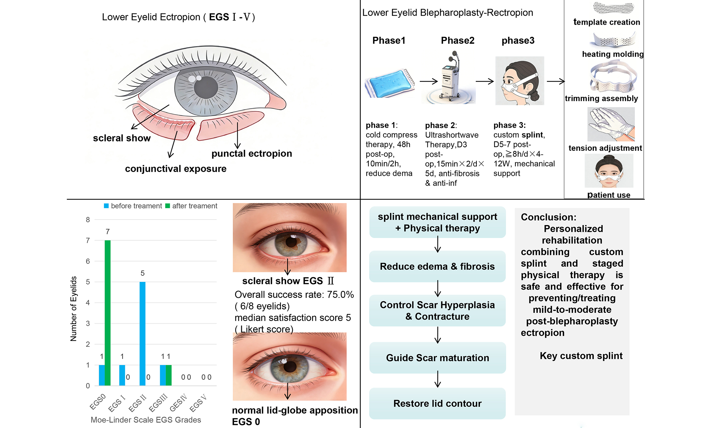
Lower eyelid ectropion is a clinically and aesthetically significant complication following blepharoplasty and periocular reconstruction. Although surgical correction is well-established, early postoperative nonsurgical interventions remain limited. This case series describes a structured rehabilitation protocol integrating a custom-fabricated low-temperature thermoplastic eyelid splint with targeted physical therapy for early correction and prevention. We enrolled six patients (eight eyelids) with postoperative lower eyelid ectropion or at high risk for its development in a custom rehabilitation protocol comprising the splint and a structured physical therapy regimen of immediate cold-compress therapy followed by ultrashortwave therapy. Outcomes were evaluated using the validated Moe-Linder ectropion grading scale (EGS), symptom resolution, and patient-reported satisfaction (5-point Likert scale); safety was monitored by recording splint-related adverse events. At 6-month follow-up, among seven therapeutic eyelids, six improved to EGS grade 0, while the prophylactic eyelid maintained grade 0, totaling 7/8 eyelids at grade 0. Of these, five had complete symptom resolution, whereas one showed partial dry eye improvement. One eyelid with EGS grade III ectropion remained grade III. No splint-related complications occurred. Median satisfaction was 5 (range: 4-5), with 7/8 eyelids scoring 5. Early rehabilitation with a custom thermoplastic splint and physical therapy appears effective and safe for postoperative lower eyelid ectropion, particularly in mild to moderate cases, while also serving as a prophylactic measure and offering adjunctive benefits in selected complex cases, though its efficacy appears limited in severe (EGS grade III) ectropion.
Keywords
INTRODUCTION
Lower eyelid ectropion is a challenging complication following procedures such as blepharoplasty, tumor excision, and traumatic reconstruction. Its pathogenesis involves a disruption of eyelid architecture and tension balance, including anterior lamellar shortage, vertical tension gradient abnormalities, horizontal laxity, and postoperative edema[1-5]. This malposition manifests as lid retraction, punctal eversion, conjunctival exposure, epiphora, photophobia, and dry eye, which, if prolonged, can lead to corneal pathology, recurrent infection, and chronic ocular surface damage[6].
The primary therapeutic goal is to restore the physiological eyelid-globe relationship. Current postoperative measures, however, have significant limitations. Frost sutures provide temporary vertical support but may lose effectiveness over time[7,8]; Long-term use of double eyelid tape may lead to eyelid laxity, adversely affect ocular health, and complicate blepharoplasty procedures[9]; massage techniques require sustained patient compliance with variable efficacy, particularly in moderate-to-severe cases[10]; and while cold compress therapy with head elevation reduces edema, it does not actively correct malposition. The common limitation of these approaches is their inability to provide sustained, adjustable mechanical support to guide tissue healing, which may explain why some early ectropion cases progress to require surgical correction.
While surgical techniques - including lateral canthopexy, skin grafting, spacer grafts, and midface lifting - are well-established[11-14], standardized nonsurgical rehabilitation strategies capable of interrupting ectropion progression in the early postoperative period remain scarce. To address this gap, we developed an integrated rehabilitation protocol combining staged physical therapy (cold compress therapy followed by ultrashortwave therapy) with a custom low-temperature thermoplastic eyelid splint. This combination is designed to simultaneously address inflammatory edema and provide precise, adjustable mechanical support to guide tissue contour during healing. In this case series, we report the clinical feasibility, safety, and short-term outcomes of this protocol in six patients (including three representative cases).
MATERIALS AND METHODS
Patient selection
This retrospective case series included six consecutive patients (eight eyelids) treated with our custom low-temperature thermoplastic splint protocol between January 2022 and December 2023. Of these, one eyelid received prophylactic treatment following congenital melanocytic nevus excision, and seven eyelids received therapeutic treatment for established ectropion, including one case following traumatic wound repair, three cases following previous failed ectropion correction surgery, two cases following bilateral lower blepharoplasty performed at external institutions, and one case following post-traumatic lower eyelid reconstruction. Patients were included if they either (1) presented with postoperative ectropion following blepharoplasty or periocular reconstruction; or (2) were deemed at high risk for ectropion development, warranting prophylactic intervention. Notably, three patients (four eyelids) had undergone prior unsuccessful surgical repair, indicating complex pathology. All patients provided written informed consent for the publication of clinical photographs. Demographic and clinical characteristics, including ectropion severity graded by the validated Moe-Linder ectropion grading scale (EGS)[12], are summarized in Tables 1 and 2.
Demographic and clinical characteristics of study patients
| Case | Sex | Age (year) | Affected side | Previous surgery | Preoperative diagnosis | Current surgery | Indication | Baseline EGS (L/R) | Final EGS at 6 Mon (L/R) |
| Case 1 | Male | 38 | Right | Debridement and suture of facial soft tissue injury | Right facial depression deformity + right lower lid ectropion + right infraorbital sinus tract + multiple fractures | Correction of right lower lid ectropion + lower lid reconstruction + resection and closure of right infraorbital sinus tract | Established ectropion | 0/III | 0/III |
| Case 2 | Male | 60 | Bilateral | Bilateral lower blepharoplasty | Unclear (surgery at another hospital) | None | Established ectropion | I/II | 0/0 |
| Case 3 | Male | 12 | Right | None | Congenital melanocytic nevus | Resection of congenital melanocytic nevus | Prophylaxis | 0/0 | 0/0 |
| Case 4 | Female | 30 | Left | Debridement and suture of facial soft tissue injury | No abnormality (surgery at another hospital) | None | Established ectropion | II/0 | 0/0 |
| Case 5 | Female | 43 | Left | Resection of benign left lower lid mass | Left lower lid ectropion deformity + post-surgical scar contracture | Left auricular cartilage harvest + acellular dermal matrix implantation + correction of left lower lid scar contracture | Established ectropion | II/0 | 0/0 |
| Case 6 | Female | 61 | Bilateral | Bilateral lower blepharoplasty | Bilateral lower lid retraction with lateral canthal scar adhesion | Correction of bilateral lower lid ectropion/retraction + acellular dermal matrix implantation + lysis of lateral canthal scars | Established ectropion | II/II | 0/0 |
Summary of splint usage data for each case
| Case | Timing initiation | Total duration | Daily wear (h/day) | Patient adherence | Patient satisfaction | Follow-up (months) | Outcome | Complications |
| Case 1 | POD 7 | 12 weeks | 8 h/day | Good, completed | 5 | 1/3/6 | EGS grade III | None |
| Case 2 | POD 33 | 5 weeks | 12 h/day | Good, completed | 5/5 | 3/6 | Complete resolution | None |
| Case 3 | POD 7 | 12 weeks | 10 h/day | Good, completed | 5 | 1/3/6 | EGS grade maintained at 0 | None |
| Case 4 | POD 9 | 8 weeks | 10 h/day | Good, completed | 5 | 1/3/6 | Complete resolution | None |
| Case 5 | POD 7 | 12 weeks | 8 h/day | Good, completed | 5 | 1/3/6 | Complete resolution | None |
| Case 6 | POD 7 | 8 weeks | 14 h/day | Good, completed | 4/5 | 1/3/6 | Partial improvement (dry eye) | None |
Fabrication and application of the splint
The custom low-temperature thermoplastic splint was fabricated using a standardized five-step protocol [Figure 1]:
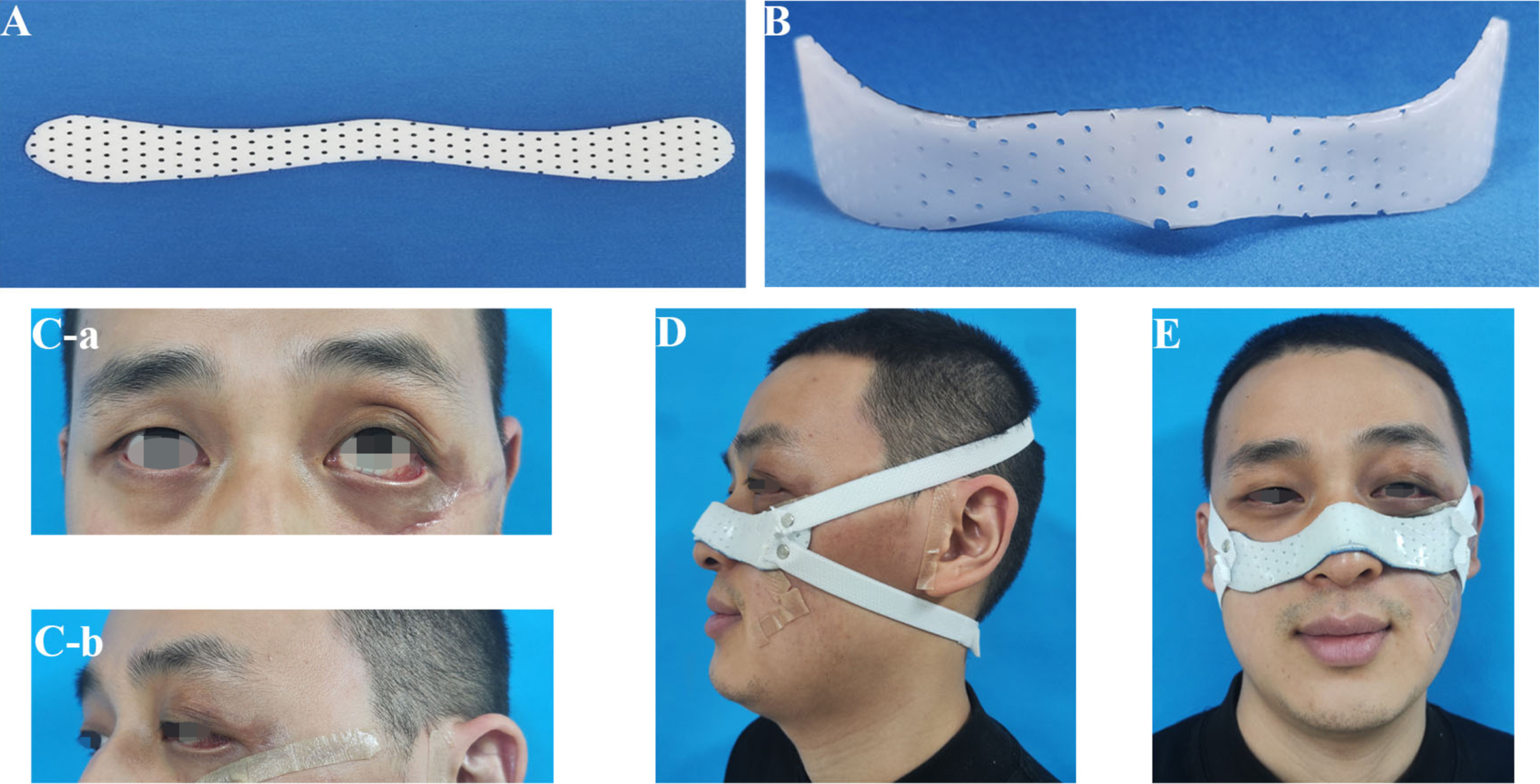
Figure 1. Steps for Fabrication and Fitting of the custom thermoplastic lower eyelid splint. (A) A paper template is trimmed according to the patient’s lower eyelid contour; (B) The thermoplastic material is softened in a 65-70 °C water bath and molded directly onto the lower eyelid to achieve precise conformity to the periorbital anatomy; (C-a and C-b) Pre-splint frontal and lateral views showing left lower eyelid ectropion; (D) Correct wearing method: the two upper straps are secured upward at an angle of approximately 45°, while the two lower straps are secured behind the ear; (E) Post-splint frontal view demonstrating upward biomechanical support and complete cosmetic correction.
Step 1 (template creation)
A paper template was created by tracing the patient’s lower eyelid contour, with adjustments made for individual anatomy. This outline was transferred to low-temperature thermoplastic sheeting (Medical Polymer Splint, Guangzhou Kailieruidi, model P-2412, 920 mm × 610 mm, 1.0 mm thickness, selected for optimal rigidity and patient comfort) and cut to shape [Figure 1A].
Step 2 (heating and molding)
With the patient seated upright, the thermoplastic sheet was immersed in a thermostatically controlled water bath (65-70 °C) until pliable. The softened material was immediately applied to the lower eyelid and molded to conform to the periorbital contour, including the infraorbital rim, eyelid margin, and existing contour defects [Figure 1B].
Step 3 (trimming and assembly)
After cooling and hardening, the splint edges were smoothed. The device was connected to a custom adjustable elastic traction system using hook-and-loop fasteners (Single-sided Fastener, FZ-1025, 9 mm ×
Step 4 (initial tension setting by therapist)
With the patient seated upright facing a mirror, the splint was positioned. The superior elastic straps (commercially available, brand Yingju, 1.5 cm × 10 m) were secured over the scalp (approximately 45° to horizontal), and inferior straps were secured behind the ears [Figure 1D]. After suture removal on day 7, the custom low-temperature thermoplastic splint was applied to achieve complete cosmetic correction of ectropion (restored lid-globe apposition, no scleral show) [Figure 1E].
Step 5 (patient instruction and daily use)
Tension was not standardized by fixed numerical values but by two reproducible clinical criteria: (1) immediate restoration of the lower eyelid margin to the normal anatomical position (EGS grade 0) with no scleral show upon splint application; and (2) absence of skin pressure injury, significant pain, or discomfort 30 min after application. When both criteria were met, the traction force was considered standardized. The splint was custom-molded to the patient’s eyelid contour and secured with elastic straps and hook-and-loop fasteners, enabling flexible, individualized tension adjustment without specialized equipment. At each follow-up visit, the splint was re-molded by heating to maintain optimal fit, and traction was adjusted according to the recovery of eyelid position. Once the correct tension was established, reference marks were placed on the straps. Patients were thoroughly instructed on proper “donning and doffing”, how to reproduce the marked tension using mirror feedback, and the requirement of at least 8 h of daily wear (primarily during sleep). They were advised to maintain the tension that achieved full correction while ensuring comfort and absence of skin blanching or pain, and to return for reassessment if correction was lost or skin irritation occurred.
Structured rehabilitation protocol
All patients followed a standardized multimodal protocol, as detailed in the timeline presented in Figure 2.
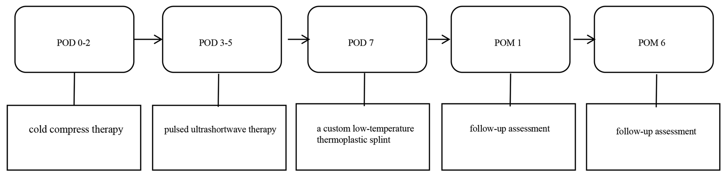
Figure 2. Detailed timeline of the rehabilitation protocol.
Phase I (cold compress therapy)
Intermittent periocular cold compress therapy (10 min every 2 h) was administered within 48 h postoperatively to control edema and mitigate inflammation.
Phase II (ultrashortwave therapy)
Beginning on postoperative day 3, ultrashortwave therapy was applied to promote tissue healing and reduce fibrosis. The treatment was delivered using a YK-C-I ultrashortwave diathermy device (Anhui Kangda Intelligent Technology Co., Ltd., China) with an operating frequency of 40.68 MHz and adjustable output power set at the lowest level (20 W) of the 20-50 W adjustable range. The device operated in continuous wave mode. Two capacitive electrodes were placed juxtaposed over both closed eyelids, with a 3-6 cm medical gauze pad used as a cushion between the electrodes and the skin for ocular protection, at an electrode-to-skin distance of approximately 2-3 cm. Each session lasted 15 min, and the treatment was administered twice daily for 5 days. Contraindications to treatment included: (1) implanted cardiac pacemaker, internal metallic foreign bodies, or heart valve replacement; (2) active pulmonary tuberculosis; (3) cardiovascular insufficiency; (4) bleeding tendency or coagulopathy; (5) malignant tumors; and (6) pregnancy (lower abdomen). Treatment was discontinued routinely upon completion of the prescribed course or achievement of significant improvement in inflammation, edema, or pain, and was terminated prematurely if the patient developed significant discomfort, worsening of symptoms, adverse reactions, or if equipment malfunction occurred.
Phase III (orthotic management)
Initiated after acute edema resolution (postoperative days 5-7). Biomechanical objectives included: (1) sustained upward vector support against gravitational and cicatricial forces; (2) gentle, consistent pressure to ensure skin-muscle flap adherence; and (3) guided scar maturation along the natural lid contour. The splint was worn for ≥ 8 h daily (primarily during sleep) for 4-12 weeks, with adjustments based on individual scar plasticity and clinical response.
A detailed timeline
The detailed timeline of the phased rehabilitation protocol is presented below [Figure 2].
Outcome measures
Assessments were performed at baseline (before splint application) and at the 6-month follow-up. The primary outcome was the change in ectropion severity graded by the EGS. EGS grading was performed independently by two senior plastic surgeons (Xi Yuan and Jie Shen) based on standardized clinical photographs taken under uniform conditions; photographs were reviewed independently, and disagreements were resolved by consensus. The assessors were not masked to treatment status due to the retrospective design of this study. Secondary outcomes included patient-reported symptom relief (epiphora, ocular irritation, xerophthalmia), overall satisfaction (assessed using a 5-point Likert scale), and safety monitoring for splint-related complications (skin breakdown, pressure injury).
RESULTS
Treatment outcomes at 6-month follow-up
At the 6-month follow-up, outcomes were stratified by treatment intent. Among the eight eyelids, six therapeutic eyelids with pre-existing ectropion improved to EGS grade 0, yielding an overall cosmetic success rate of 75.0% (6/8), or 85.7% (6/7) when the prophylactic case was excluded, while the single prophylactic eyelid maintained EGS grade 0 from baseline throughout, resulting in a total of 7/8 eyelids at EGS grade 0. Of these six cosmetically successful therapeutic eyelids, five (83.3%) reported complete resolution of ectropion-related symptoms, whereas one (16.7%) showed only partial improvement in dry eye symptoms. The single prophylactic eyelid (baseline EGS grade 0) maintained normal eyelid position, contour, and function throughout the follow-up period. One therapeutic eyelid (Case 1) with EGS grade III ectropion following post-traumatic lower eyelid reconstruction did not show significant improvement and remained EGS grade III at the final follow-up.
Baseline characteristics
Before initiation of this physical therapy, the distribution of ectropion severity according to the EGS grading system was as follows: one eyelid (prophylactic application following congenital melanocytic nevus excision) was graded as EGS grade 0; one eyelid (left eye, ectropion secondary to traumatic wound repair at an external institution) was graded as EGS grade I; one eyelid (right eye, ectropion following traumatic wound repair at an external institution) was graded as EGS grade III (Case 1); and the remaining five eyelids were graded as EGS grade II. Among the EGS II group, one patient who underwent lower blepharoplasty at an external institution had bilateral lower eyelid ectropion, with EGS grade II ectropion in the right eye; one patient with left lower eyelid ectropion remained at EGS grade II on postoperative day 7 after revision surgery; one patient developed bilateral EGS grade II ectropion following lower blepharoplasty at an external institution; and one patient developed EGS grade II ectropion in the left eye following debridement and suture of facial soft tissue injury at an external institution.
Patient satisfaction and safety
Satisfaction was assessed for each treated eyelid using a 5-point Likert scale at 6 months. Among the eight eyelids, seven (87.5%) scored 5 (very satisfied), and one (12.5%) scored 4 (satisfied) because of incomplete resolution of dry eye symptoms, yielding a median score of 5 (range: 4-5). No splint-related complications were observed throughout the follow-up period.
Representative cases
The protocol described in the Methods section represents the recommended standardized treatment regimen. In clinical practice, two patients (Cases 2 and 4) were referred from external institutions and did not receive the standardized protocol within the recommended time windows; they were referred to our department and subsequently received ultrashortwave therapy combined with splint application upon referral. Case 2 has been described in detail in Section “Case 2: eyelid malposition following blepharoplasty” as a representative example of this scenario. Deviations from the recommended protocol for all cases are summarized in Table 2.
Case 1: cicatricial ectropion following severe facial trauma reconstruction
A 38-year-old man was referred for rehabilitation following severe periorbital trauma sustained in a fall. After initial debridement and suturing of the right face, he developed a right facial depression deformity, a severe cicatricial ectropion (EGS grade V) of the right lower eyelid, and an infraorbital sinus tract formation [Figure 3A]. The patient underwent a complex reconstructive procedure, including correction of lower eyelid ectropion, reconstruction of the right lower eyelid, and excision and closure of the infraorbital sinus tract. A 2 cm × 4 cm tongue-shaped flap was designed in the right lateral orbital region, elevated, and rotated 90° to repair the lower eyelid defect. Concurrently, a 1 cm × 2 cm auricular cartilage graft was harvested and trimmed into a π-shaped strut, which was fixed to the periosteum of the inferior orbital rim to provide lower eyelid support reconstruction. At the baseline assessment for our rehabilitation protocol on postoperative day 7 (after suture removal, before application of the custom low-temperature thermoplastic splint), the right lower eyelid exhibited a residual EGS grade III cicatricial ectropion with conjunctival edema [Figure 3B]. On postoperative day 7, the splint component of the standardized multimodal rehabilitation protocol was initiated [Figure 3C]; cold compress therapy had been administered on POD 0-2, and ultrashortwave therapy on POD 3-7. Splint application provided immediate mechanical support by reducing downward traction and applying mild pressure to the skin flap, thereby improving lid-globe apposition. After 8 weeks of consistent daily use (8 h/day), mild swelling of the lower eyelid was observed [Figure 3D], and the patient continued splint use until the full 12-week protocol was completed. At the 6-month postoperative visit, although EGS grade III ectropion with mild scleral show persisted objectively, satisfactory eyelid contour was achieved and the flaps and scars healed without complications. The patient reported being satisfied with the aesthetic outcome. Favorable scar control was observed during the period of sustained mechanical off-loading provided by the splint, which contributed to stable flap and scar healing without contracture recurrence [Figure 3E].
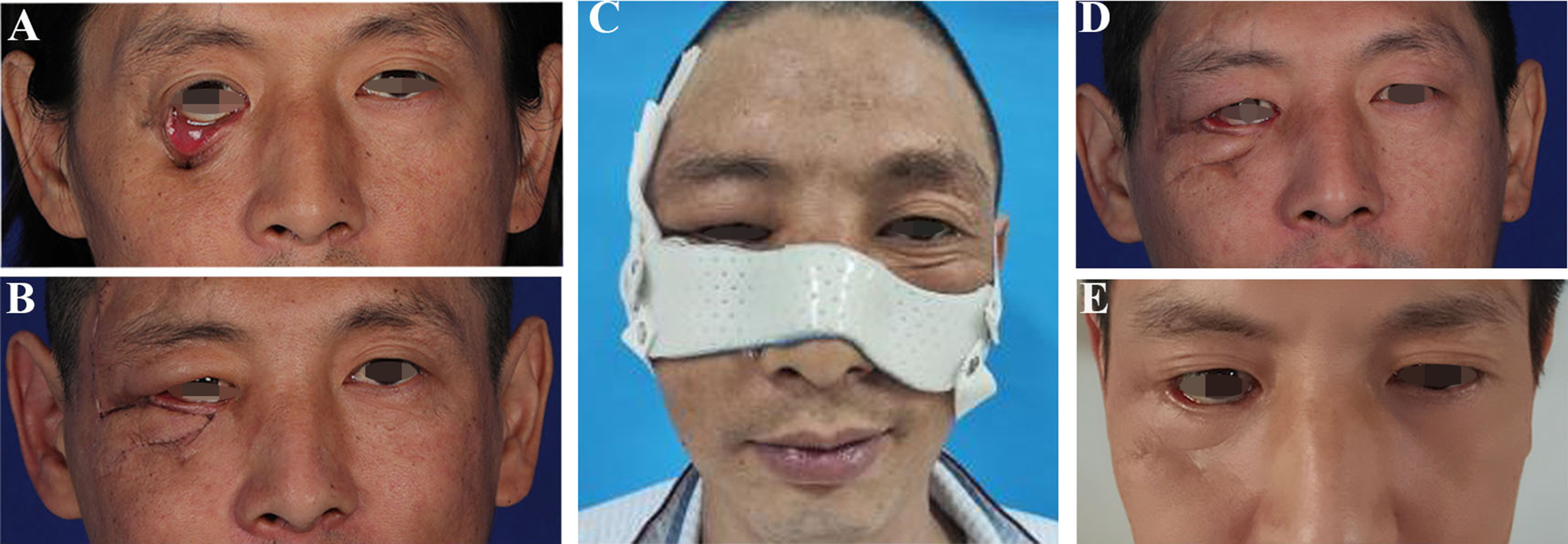
Figure 3. Surgical correction of cicatricial lower eyelid ectropion in a 38-year-old male patient. (A) Preoperative view showing severe right periorbital trauma with tissue loss and a midface defect (30 mm × 20 mm). EGS grade V ectropion prior to definitive reconstructive surgery, with complete conjunctival exposure, punctal eversion, and lagophthalmos; (B) Baseline status on postoperative day 7 (after suture removal, before application of the custom low-temperature thermoplastic splint) following reconstructive surgery, demonstrating persistent EGS grade III ectropion with conjunctival edema; (C) Status immediately after splint application on postoperative day 7 showing immediate improvement in lid-globe apposition; (D) At the 8-week interim assessment, mild swelling of the lower eyelid was observed; (E) At the 6-month postoperative visit, although EGS grade III ectropion with mild scleral show persisted objectively, satisfactory eyelid contour was achieved and the flaps and scars healed without complications. The patient reported being satisfied with the aesthetic outcome. EGS: Ectropion grading scale.
Case 2: eyelid malposition following blepharoplasty
A 60-year-old male patient developed lower eyelid ectropion following cosmetic lower blepharoplasty performed at an external institution and was referred to our department on postoperative day 33. The patient reported having received local cold compress therapy within the first 48 h after surgery at the referring institution, but conservative management had failed. Examination on presentation revealed significant horizontal laxity of the lower eyelid. According to the Moe-Linder Grading Scale, the right eyelid was graded EGS grade II (scleral show) and the left eyelid was graded EGS grade I (mild scleral show) [Figure 4A and B]. Intervention commenced on the same day. The standardized multimodal rehabilitation protocol - comprising ultrashortwave therapy and a custom low-temperature thermoplastic splint - was initiated [Figure 4C]. The splint was employed to provide superolateral support. After 5 weeks of compliant use, both eyelids achieved EGS grade 0 (normal position and function). At the 6-month follow-up, cosmetic correction was maintained, with complete resolution of all symptoms [Figure 4D].
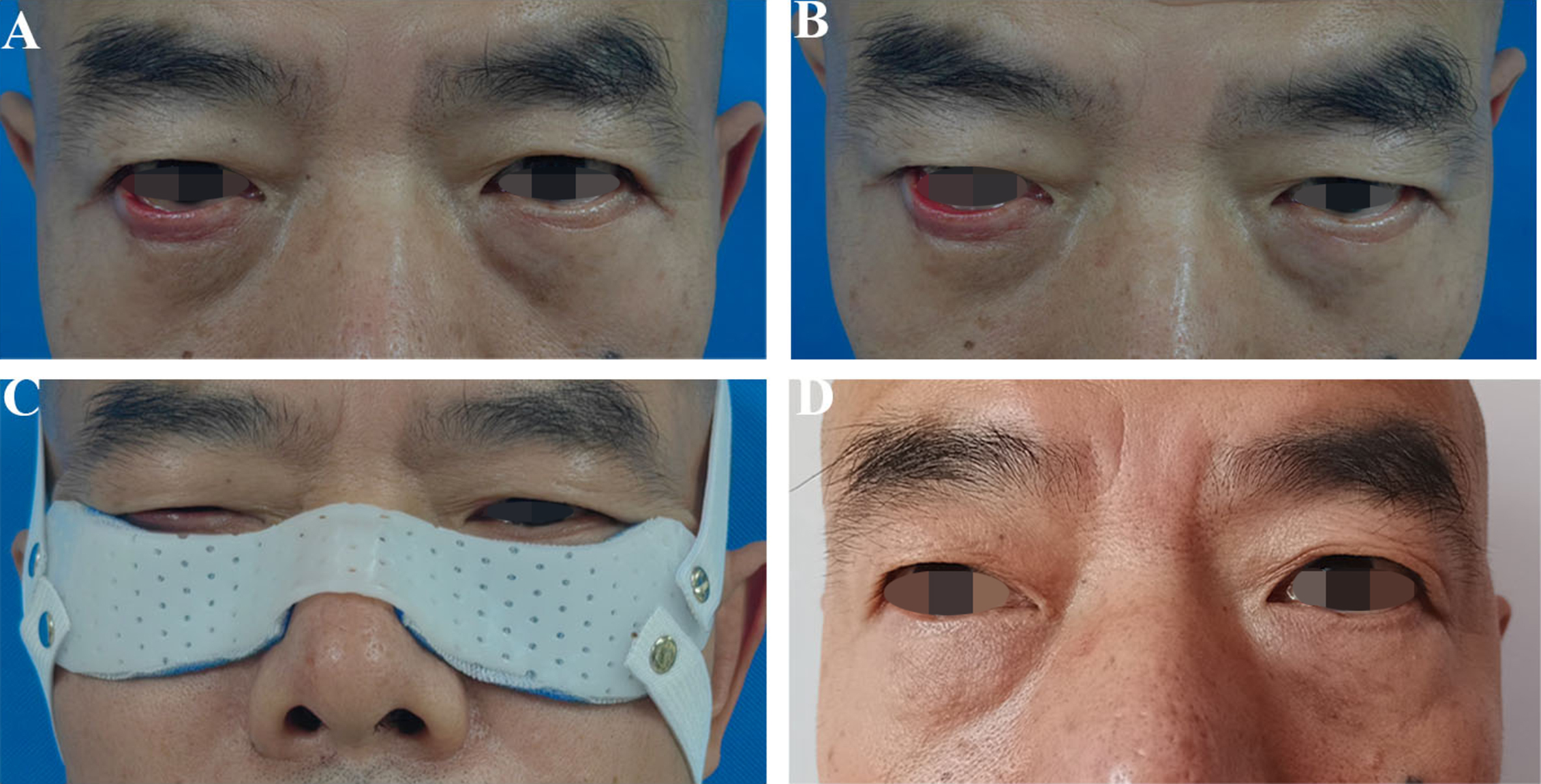
Figure 4. Correction of postoperative lower eyelid ectropion in a 60-year-old male patient. (A and B) Bilateral lower eyelid malposition following cosmetic blepharoplasty. Pre-intervention assessment (postoperative day 33) showing horizontal laxity, with EGS grade I (mild scleral show) malposition on the left and EGS grade II (scleral show) on the right; (C) The patient wearing the custom low-temperature thermoplastic splint; (D) Appearance at the 6-month follow-up, demonstrating restoration of normal eyelid position and complete resolution of symptoms. EGS: Ectropion grading scale.
Case 3: prophylactic application after congenital nevus excision
A 12-year-old boy underwent excision of a congenital melanocytic nevus (19 mm × 12 mm) on the right lower eyelid [Figure 5A]. A vertical auxiliary incision was required for wound closure, creating a high risk for cicatricial ectropion due to anticipated vertical scar contracture. To prevent this complication, a tongue-shaped flap with a pedicle width of 1.2 cm and a length of approximately 2.5 cm was designed in the right nasofacial groove to cover the right lower eyelid defect [Figure 5B]. The full integrated protocol was initiated immediately after surgery, comprising cold-compress therapy on postoperative days 0-2, ultrashortwave therapy on postoperative days 3-7, and custom splint application on postoperative day 7. The splint was applied specifically to provide gentle, sustained superolateral support, aiming to neutralize contractile forces and guide scar remodeling [Figure 5C]. Following 8 weeks of compliant splint use (10 h/day), edema had subsided without any signs of ectropion development, with EGS grade 0 maintained [Figure 5D], and the patient continued splint use until the full 12-week protocol was completed. At the 6-month follow-up, normal eyelid position, contour, and function were maintained, confirming successful prevention of ectropion [Figure 5E].
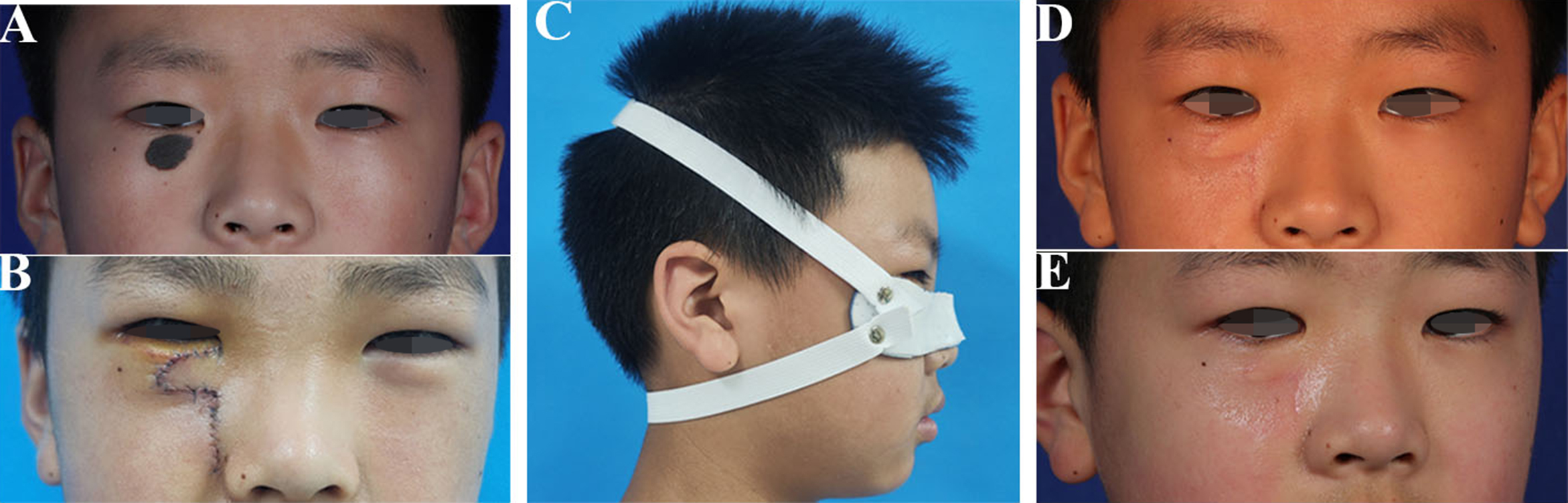
Figure 5. Comprehensive management of a congenital melanocytic nevus of the right lower eyelid in a 12-year-old male patient. (A) Preoperative appearance of a congenital melanocytic nevus (19 mm × 12 mm) on the right lower eyelid; (B) Intraoperative view after nevus excision and local flap reconstruction, highlighting the vertical (auxiliary) incision that conferred a high risk for cicatricial ectropion; (C) The custom low-temperature thermoplastic splint was applied on postoperative day 7 as part of the prophylactic protocol; (D) At the 8-week interim assessment, resolution of edema and controlled scar remodeling were observed without ectropion, with EGS grade 0 maintained; (E) Outcome at 6-month follow-up, demonstrating maintained normal eyelid position, contour, and function, confirming successful prevention of ectropion. EGS: Ectropion grading scale.
DISCUSSION
This case series demonstrates that a multimodal rehabilitation protocol integrating physical therapy with a custom biomechanical splint may effectively alter the early postoperative trajectory of lower eyelid ectropion. The clinical efficacy is attributable to several interconnected pathophysiological mechanisms.
Early postoperative ectropion is driven by transient yet potent biomechanical forces: downward traction from edema overwhelming weakened orbicularis support[4]; vertical lamellar mismatch from anterior lamellar shortening[2,3]; horizontal canthal laxity increasing lateral eversion risk[15]; and progressive anterior lamellar contracture from scar proliferation[13,16]. If not addressed in a timely manner, these forces culminate in permanent malposition.
Mechanism of action of ultrashortwave therapy
Ultrashortwave therapy exerts its core therapeutic effects through electromagnetic field-induced thermal and non-thermal actions. The underlying mechanisms involve modulation of macrophage polarization and inflammatory signaling pathways to reduce local inflammatory mediator levels, as well as regulation of fibroblast activity and collagen deposition under specific parameters[17,18]. These effects collectively achieve anti-inflammatory responses, promotion of tissue repair, and inhibition of abnormal scar formation. However, the therapeutic efficacy is dose-dependent, and excessive intensity may paradoxically induce pro-inflammatory and pro-fibrotic reactions[18]. Ultrashortwave therapy was not employed as an independent treatment for ectropion, but rather as part of a comprehensive rehabilitation protocol, working synergistically with the mechanical support provided by the splint. These actions collectively reduce edema and abnormal interlamellar tension, thereby mitigating the primary downward traction forces that contribute to lower eyelid ectropion.
Biomechanical advantages of the custom splint
The custom splint provides active biomechanical regulation with distinct advantages: precise cosmetic conformity, balanced bidirectional elastic vectors, sustained pressure for scar management, enhanced flap adherence reducing hematoma/seroma risk, and dynamic adjustability as edema subsides. The superolateral vector specifically counteracts both gravitational pull and cicatricial contracture, while the conformational pressure guides scar maturation along the physiological lid contour.
Clinical implications
In clinical practice, although Case 2 did not strictly comply with the 48-h protocol initiation window, the favorable outcome suggests that subsequent interventions (ultrashortwave therapy and splint application) may still provide therapeutic benefit for established ectropion, even when the early cold compress therapy window has been missed. This protocol is intended as an adjunctive treatment modality for two patient categories: (1) prophylactic application in high-risk patients (e.g., congenital melanocytic nevus excision, traumatic reconstruction) where early intervention mitigates scar traction and provides long-term stability; and (2) therapeutic application in mild-to-moderate ectropion (EGS grades I-II), where early mechanical support combined with anti-inflammatory therapy reverses malposition before fibrosis develops. This protocol is particularly suitable for patients at risk of malar edema, those predicted to develop postoperative ectropion, and those requiring temporary support without invasive procedures. Our preliminary experience validates its feasibility and efficacy.
However, clinical application demonstrated that one eye undergoing post-traumatic cicatricial ectropion correction maintained EGS grade III ectropion of the right lower eyelid at the 6-month follow-up [Figure 3E], despite the absence of corneal irritation symptoms and high patient satisfaction. The limited therapeutic efficacy can be attributed to the following factors. First, primary trauma and scar formation: the patient sustained multiple facial fractures and soft tissue contusions following a fall. After debridement and primary closure at an external institution, cicatricial contracture of the anterior lamella (skin and orbicularis oculi muscle) developed, resulting in lagophthalmos and severe lower eyelid ectropion (EGS grade V), with complete tarsal plate exposure and conjunctival epithelialization. Pre-existing soft tissue defects, severe scarring, and insufficient tissue volume collectively constituted a high-risk substrate for ectropion formation before the current reconstruction. Second, local tissue disruption and sinus tract involvement: a 3 cm × 2 cm facial depression with a sinus tract was present in the right infraorbital region. During the reconstructive procedure, incision of the skin and orbicularis oculi muscle with dissection along the sinus tract further disrupted the local anatomical architecture. The subsequent healing process across multiple tissue layers was compromised, significantly increasing the risk of lower eyelid ectropion recurrence. This underscores the limited corrective capacity of nonsurgical approaches in severe cases with extensive soft-tissue deficits, deep structural disruption, and graft contracture. In such scenarios, surgical reconstruction remains indispensable, and our protocol may serve as a useful adjunct to consolidate surgical outcomes and inhibit scar reformation.
Strengths and limitations
To our knowledge, this is the first study to introduce a systematic postoperative rehabilitation pathway integrating local physical therapy modalities with a precise biomechanical splint. The protocol unifies three core objectives - cosmetic restoration, complication prevention, and scar management - within a single framework. The custom splint effectively counteracts vertical gravitational pull while ensuring patient comfort and compliance. Its potential influence on concomitant horizontal laxity warrants further investigation. Preliminary efficacy across varied etiologies suggests potential applicability in similar contexts.
However, we acknowledge several limitations in this study. First, the single-center retrospective design with a small sample size (n = 6) limits the generalizability of our findings. Second, the absence of a concurrent control group and factorial design precludes differentiation of the individual effects of ultrashortwave therapy, splint application, or their combination. Third, the assessors were not masked to treatment status due to the retrospective nature of the study, and inter-rater reliability was not formally assessed using a statistical measure owing to the small sample size; these factors should be considered when interpreting the EGS-based outcomes. Fourth, we acknowledge the lack of data on the overall incidence of post-blepharoplasty ectropion at our institution as a limitation. Future factorial randomized controlled trials are needed to isolate each component’s independent contribution, determine the optimal regimen across clinical settings, and validate our preliminary findings. Notably, our current protocol does not include Hilotherapy or hyaluronidase-based therapy - both of which have demonstrated efficacy in specific edema contexts (early postoperative swelling and HA-related refractory edema, respectively)[19,20]. Future studies may therefore investigate the potential value of integrating these modalities into the present rehabilitation protocol or compare their relative effectiveness.
CONCLUSIONS
In this technical note, we describe and preliminarily evaluate an integrated, nonsurgical rehabilitation protocol for managing and preventing lower eyelid ectropion following various periocular procedures, combining cold compress therapy, ultrashortwave therapy, and a custom low-temperature thermoplastic splint. The protocol is designed to address postoperative inflammation and provide mechanical support to the lower eyelid through staged, timed interventions. In our preliminary case series, this approach was safe, feasible, and well-tolerated. It appeared to be effective in preventing ectropion and in correcting early mild-to-moderate cases. In selected severe cicatricial ectropion, it may serve as a useful adjunct to support scar remodeling and improve patient comfort, although its corrective efficacy remains limited. While our findings suggest that this protocol may represent a promising strategy for ectropion management, we acknowledge that the current evidence is preliminary and based on a limited case series. Long-term outcomes and definitive efficacy require validation through multicenter, controlled trials with larger sample sizes.
DECLARATIONS
Authors’ contributions
Responsible for designing the research protocol and participated in manuscript review: Jiang X, Zhang J
Performed clinical treatments and drafted the manuscript: Gao L
Involved in the collection of clinical data: Li K, Lei W
Took charge of the design and implementation of surgical protocols: Yuan X, Shen J
All authors have read and approved the final version of the manuscript.
Availability of data and materials
Not applicable.
AI and AI-assisted tools statement
During the preparation of this manuscript, the AI tool Doubao (version 2.0, released 2026-02-14) was used solely for language editing, grammar checking, and translation. These tools did not influence the study design, data collection, analysis, interpretation, or the scientific content of the work. All authors take full responsibility for the accuracy, integrity, and final content of the manuscript.
Financial support and sponsorship
This research was supported by the Clinical Innovation Technology Cultivation Program of Southwest Hospital (2025CXJS19) and Chongqing Science and Health Joint General Project (2025MSXM082).
Conflicts of interest
All authors declared that there are no conflicts of interest.
Ethical approval and consent to participate
This study was approved by the Ethics Committee of the First Affiliated Hospital of Army Medical University of the Chinese People’s Liberation Army [Approval No. (B2) 2025KY037]. The study was conducted in accordance with the principles of the Declaration of Helsinki, and all research procedures complied with relevant medical ethics standards. As this study was a retrospective analysis of previously collected clinical data, the requirement for informed consent for the retrospective use of clinical data was waived by the ethics committee.
Consent for publication
Written informed consent for the publication of clinical information and identifiable clinical photographs was obtained from all patients. For patients who were unable to provide consent personally, written informed consent for publication was obtained from their legal guardians.
Copyright
© The Author(s) 2026.
REFERENCES
1. McCord CD Jr, Shore JW. Avoidance of complications in lower lid blepharoplasty. Ophthalmology. 1983;90:1039-46.
2. Harounian J, Wulc AE, Brackup AB, Ramesh S. Subtle eyelid retraction after lower blepharoplasty. J Plast Reconstr Aesthet Surg. 2019;72:1682-7.
3. Steinsapir KD, Steinsapir S. The treatment of post-blepharoplasty lower eyelid retraction. Facial Plast Surg Clin North Am. 2021;29:291-300.
4. Cheng T, Yang B, Zhang Y, Tang S. Temporary ectropion caused by blepharoplasty for the lower eyelid: a case report. Asian J Surg. 2021;44:1186-8.
6. Bergstrom R, Czyz CN. Ectropion lower eyelid reconstruction. In: StatPearls [Internet]. Treasure Island (FL): StatPearls Publishing; 2026.
7. Connolly KL, Albertini JG, Miller CJ, Ozog DM. The suspension (Frost) suture: experience and applications. Dermatol Surg. 2015;41:406-10.
8. Yalcin CE. Holding the lid: suspension techniques to prevent lower eyelid ectropion-a systematic review. Facial Plast Surg. 2026;42:451-6.
9. Yang K, Wang X, Sun Y, et al. Implications of long-term double eyelid tape use. Aesthetic Plast Surg. 2025;49:75-86.
10. Hwang SW, Hwang K. Eyelid finger manipulation in the treatment of overcorrected blepharoptosis and postblepharoplasty ectropion-retraction. Plast Reconstr Surg. 2015;136:700e.
11. Kim KH, Baek JS, Lee S, et al. Causes and surgical outcomes of lower eyelid retraction. Korean J Ophthalmol. 2017;31:290-8.
12. Moe KS, Linder T. The lateral transorbital canthopexy for correction and prevention of ectropion: report of a procedure, grading system, and outcome study. Arch Facial Plast Surg. 2000;2:9-15.
13. Mol I, Paridaens D. Efficacy of lateral eyelid-block excision with canthoplasty and full-thickness skin grafting in lower eyelid cicatricial ectropion. Acta Ophthalmol. 2019;97:e657-61.
14. Rubino C, Trignano E, Dore S, et al. 3-step combined technique for correction of involutional lower eyelid ectropion: a case series. J Clin Med. 2024;14:128.
15. Damasceno RW, Osaki MH, Dantas PE, Belfort R Jr. Involutional entropion and ectropion of the lower eyelid: prevalence and associated risk factors in the elderly population. Ophthalmic Plast Reconstr Surg. 2011;27:317-20.
16. McKelvie J, Papchenko T, Carroll S, Ng SG. Cicatricial ectropion surgery: a prospective study of long-term symptom control, patient satisfaction and anatomical success. Clin Exp Ophthalmol. 2018;46:1002-7.
17. Ning Y, Wang Y, Guo M, et al. Ultra short waves alleviate ulcerative colitis via TRPV2 mediated-macrophage polarization. iScience. 2026;29:115929.
18. Huang PP, Zhang R, Zhang XF, et al. Effects of ultrashort wave diathermy on skin wounds in rabbit ears. Connect Tissue Res. 2023;64:569-78.
19. Liu J, Fan W, Guo Y, et al. Eyelid edema reduction following blepharoplasty: a 3D imaging-based pilot study on the efficacy of Hilotherapy. Quant Imaging Med Surg. 2025;15:9170-8.
Cite This Article
 Open Access
How to Cite
Download Citation
Export Citation File:
Type of Import
Tips on Downloading Citation
Citation Manager File Format
Type of Import
Direct Import: When the Direct Import option is selected (the default state), a dialogue box will give you the option to Save or Open the downloaded citation data. Choosing Open will either launch your citation manager or give you a choice of applications with which to use the metadata. The Save option saves the file locally for later use.
Indirect Import: When the Indirect Import option is selected, the metadata is displayed and may be copied and pasted as needed.
About This Article
Special Topic
Copyright

Data & Comments
Data
0






Comments
Comments must be written in English. Spam, offensive content, impersonation, and private information will not be permitted. If any comment is reported and identified as inappropriate content by OAE staff, the comment will be removed without notice. If you have any queries or need any help, please contact us at support@oaepublish.com.