


fig2
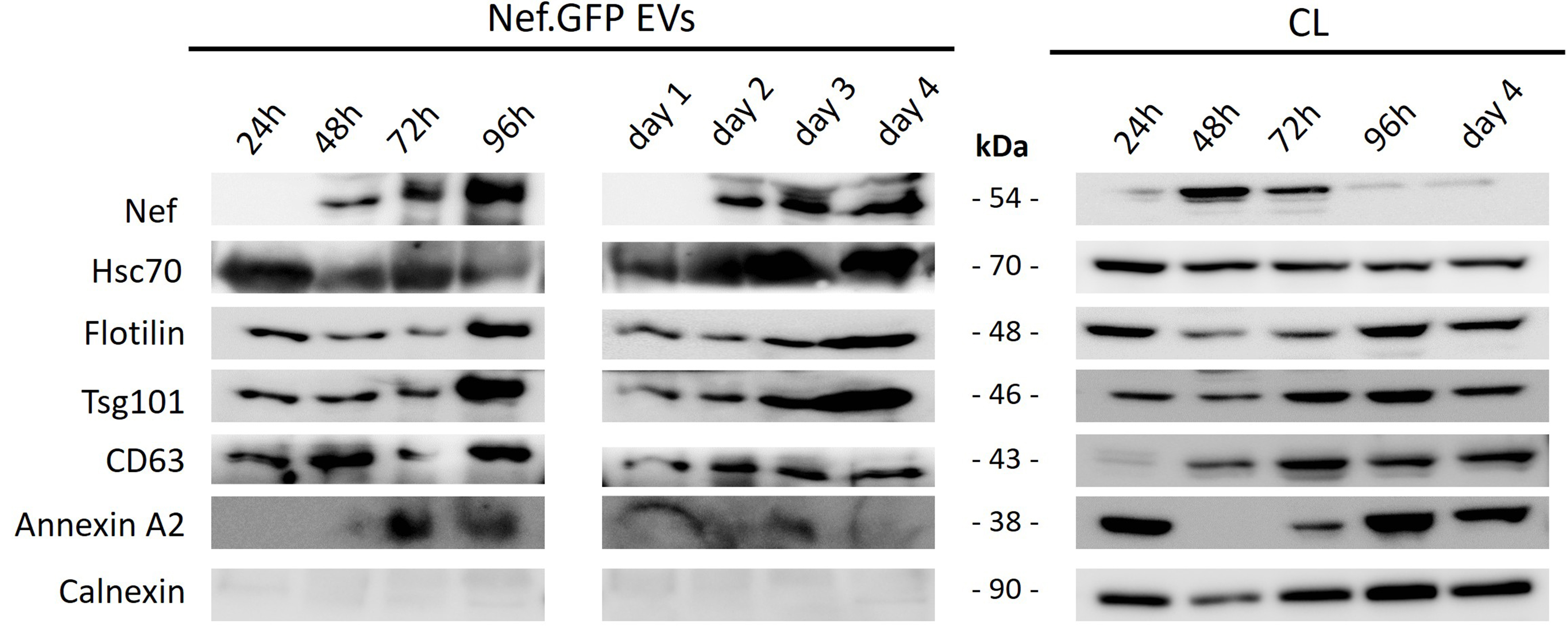
Figure 2. Continuous release of Nef.GFP-positive vesicle-like structures from h-microglia transiently expressing Nef.GFP. Immunoblot analysis of crude EVs enriched from the media of h-microglia cultures transiently expressing Nef.GFP at the indicated times, either cumulatively after 24, 48, 72, and 96 h in culture, or by sampling the same culture on days 1, 2, 3, and 4, with growth media replaced after each sampling. For samples, cells were collected at the end points in both growth experiments, that is after 24, 48, 72 and 96 h, and after day 4. Antibodies were directed against GFP, typical EV proteins (Hsc70, Flotillin, Tsg101, CD63, Annexin A2) and EV impurity marker Calnexin. CL: Cell lysate; Nef.GFP: Nef green fluorescent protein; h-microglia: human microglia; EV: extracellular vesicle; Hsc70: heat shock cognate 70; Tsg101: tumor susceptibility gene 101; CD63: cluster of differentiation 63; Annexin A2: annexin A2 protein.


