


fig1
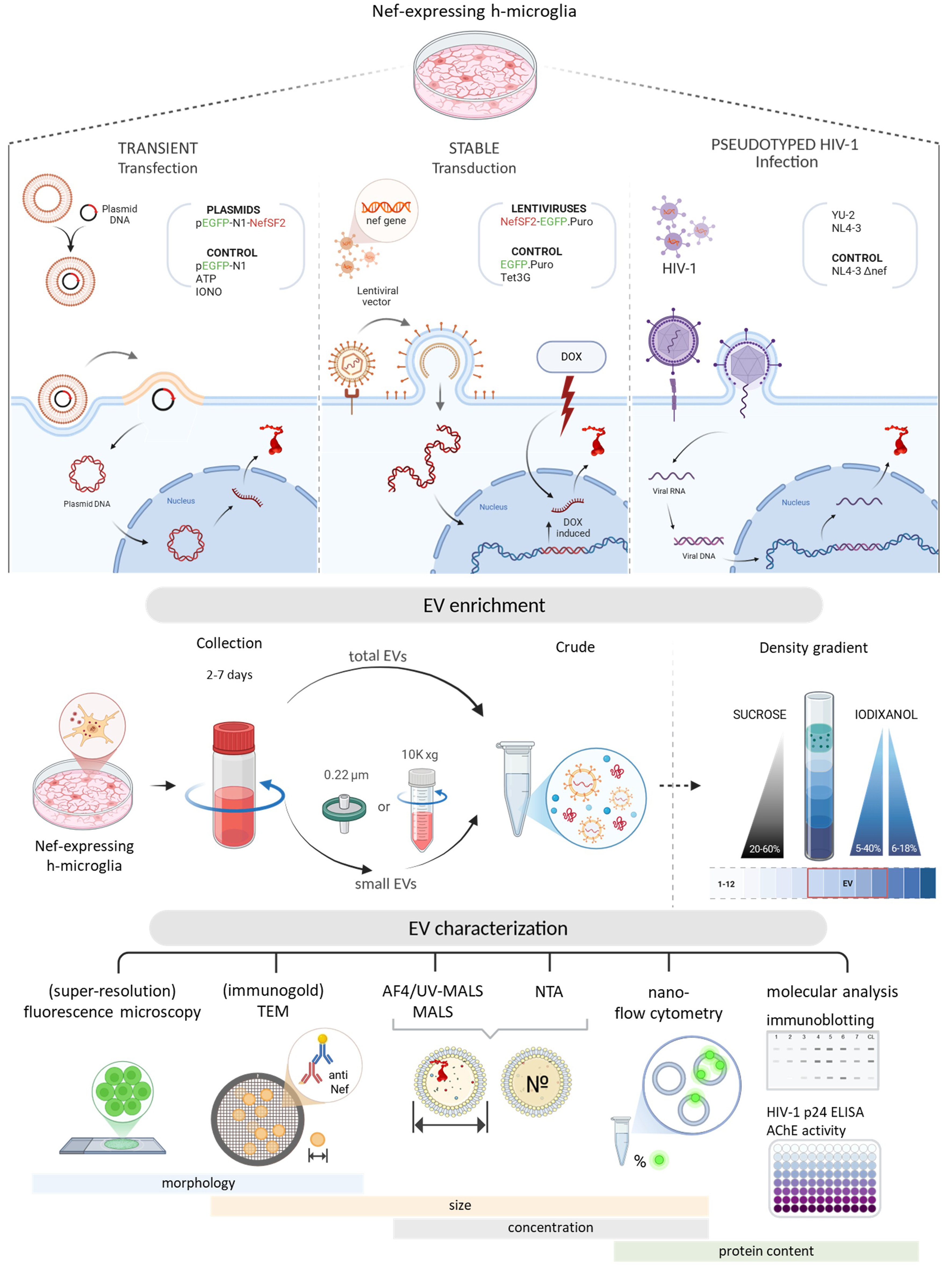
Figure 1. Schematic overview of the experimental workflow. Top panel: Three cellular models used to study the effect of Nef expression in h-microglia on vesiculation: transient expression following plasmid transfection, regulated expression from an integrated gene via lentiviral transduction, and expression from an integrated provirus after HIV-1 infection; Middle panel: Approaches for EV enrichment from conditioned media, including differential centrifugation and density gradient fractionation, to obtain total EVs or small EVs; Bottom panel: Techniques used to study EV characteristics; size, concentration and protein content were studied by (super-resolution) fluorescence microscopy, (immunogold) TEM, AF4-MALS, NTA, nano-flow cytometry, immunoblotting and other molecular approaches. DOX: Doxycycline; EV: extracellular vesicles; TEM: transmission electron microscopy; AF4-MALS: asymmetric flow field-flow fractionation coupled to a multi-angle light scattering detector; NTA: nanoparticle tracking analysis; HIV: human immunodeficiency virus.


